
Background: Ethambutol is a first‑line antituberculosis drug whose dose‑dependent optic neuropathy may lead to irreversible vision loss if undetected. However, real-world data on its ocular impact from Sudan remain scarce, despite a high tuberculosis burden and prevalent malnutrition. To describe the visual function findings (visual acuity, color vision, pupillary reactions, intraocular pressure, and fundoscopic appearance) in tuberculosis patients receiving ethambutol-containing regimens.
Methods: A hospital‑based cross-sectional study was conducted from July to December 2024. Seventy‑three adult TB patients receiving ethambutol without pre-existing vision problems were interviewed, and ocular assessments, including visual acuity, Ishihara color vision, pupil reactions, intraocular pressure, and direct fundoscopy, were performed by ophthalmologists. Data were analyzed descriptively.
Results: Most participants were 25–34 years old (42.5 %) and male (53.4 %). After commencing ethambutol, 13.7 % reported subjective vision changes, and symptoms suggestive of ocular toxicity (e.g., blurred central vision, difficulty reading) were present in 15.1 %. Despite this, 94.5 % of right eyes and 95.9 % of left eyes maintained 6/6 visual acuity, and abnormal color vision was documented in only 1.4 % of eyes. Fundoscopy revealed optic‑nerve or retinal abnormalities in 1.7 % of patients. Notably, no participant reported any chronic comorbidity, and 82.2% consumed vitamin-rich or diverse diets.
Conclusion: Clinically significant ethambutol‑induced ocular toxicity was infrequent in this Sudanese cohort, possibly due to the absence of comorbidities and favorable nutritional status.
Ethambutol, Sudan, Toxic optic neuropathy, Tuberculosis, Visual acuity, Malnutrition
Tuberculosis (TB) remains one of the world’s deadliest infectious diseases, with 10.7 million new cases reported in 2024 [1]. The most commonly recommended treatment regimen for drug-susceptible TB is 2HRZE/4HR, where rifampicin, isoniazid, pyrazinamide, and ethambutol are administered at the early stage [2]. Ethambutol hydrochloride is one of the first-line medications for tuberculosis, utilized during the initial intensive phase for categories I and II [3]. Despite its effectiveness against Mycobacterium tuberculosis, it exhibits a dose-dependent adverse effect known as Ethambutol-induced optic neuropathy (EON) [4], which often remains under-detected.
The reported incidence of EON varies considerably within the literature, ranging from 0.5% to over 35% [5]. Several factors have been reported that explain this stark variation in incidence of EON, including daily dosage, treatment duration, patient populations, nutritional status, comorbidity burden, and the sensitivity of detection methods utilized across studies [6-8]. The widely hypothesized pathogenic model of ethambutol toxicity involves disruption of oxidative phosphorylation in mitochondria within optic nerve axons. Ethambutol acts as a chelating agent, binding cellular iron and copper. In the absence of these nutrients, the activity of complexes I and IV is disrupted, resulting in optic nerve toxicity. The papillo-macular bundle of the optic nerve requires higher levels of energy, and the ethambutol-induced mitochondrial dysfunction increases the oxidative damage within the optic nerve [9].
Several risk factors predispose patients to ethambutol-related ocular toxicity, including renal diseases, longer treatment durations, older age, female sex, nutritional deficiency, liver diseases, and higher cumulative dosages [6,10,11]. Comorbidities such as diabetes mellitus, hypertension, or HIV infection increase the risk of drug-induced neurotoxicity driven by metabolic dysregulation, impaired immune function, and compromised neural repair capacity [6,7].
Clinically, ethambutol-induced ocular toxicity manifests insidiously and is typically bilateral, and though often asymmetric [12]. The most common defects of ethambutol-induced ocular toxicity are visual field defects such as central or cecocentral scotoma (blind spot affecting central vision), bitemporal hemianopsia (loss of peripheral vision in the outer halves of the visual field, dyschromatopsia (color vision deficits, typically for red and green, temporal optic disc pallor (pale appearance of the optic nerve head) [13-15]. Ethambutol-related damage to visual function is generally reversible within a few months of discontinuing ethambutol [16], while early diagnosis is also thought to help reverse the damage [8]. However, recent studies indicate that 48% of patients still experience permanent loss of visual acuity after the discontinuation of ethambutol [17]. A common finding observed across studies is that the diagnosis of ethambutol-induced ocular toxicity was made only after symptoms appeared. Early detection and prompt discontinuation of therapy are the only effective measures to halt the progression of vision loss and allow for visual recovery [18,19].
Real-world screening for ethambutol-induced ocular toxicity remains a significant limitation in clinical practice due to differences in screening practices across various healthcare systems [20], international guidelines on the prevention and early detection of EON offering inconsistent or conflicting recommendations [21,22], and the lack of consensus on standardized method or protocol for screening [8]. There are several traditional clinical assessments for ethambutol-induced ocular toxicity, such as visual acuity (Snellen chart), Ishihara color vision testing, pupillary reaction, intraocular pressure measurement, and direct fundoscopy, which are commonly used due to their cost-efficient, accessible nature without the need for sophisticated equipment [8,23,24], making them ideal for resource-limited settings. However, newer and more sensitive methods such as optical coherence tomography (OCT), visual evoked potentials (VEP), and electroretinography (ERG). These techniques can detect subclinical ethambutol-induced ocular toxicity, such as early retinal nerve fiber layer thinning, delayed conduction, and electrophysiological abnormalities even before abnormal traditional assessment findings [8,25,26]. The downside of these technologies, such as high equipment and maintenance costs and specialized technical training, limits their accessibility, particularly in low-resource settings.
In 2023, the national incidence rate of TB in Sudan is estimated at 134 cases per 100,000 population [27], and it has long been classified as a high-burden country by WHO, contributing 11-16% cases of tuberculosis in the Eastern Mediterranean Region [28]. Prevalence of malnutrition in Sudanese TB patients is widely reported in literature, deficiency of B vitamins or micronutrients essential for optic nerve health may increase their risk of developing ethambutol-related side-effects. While ethambutol-induced ocular toxicity occurs rarely and remains significantly under-detected with serious consequences like the loss of vision, it is not routinely screened for TB patients in Sudan before or after ethambutol treatment. Furthermore, the lack of healthcare infrastructure and low resources also limit the adoption of ethambutol-induced ocular toxicity screening. Collectively, high burden of TB, lack of screening, malnutrition, and low-resource settings raise a serious concern regarding the prevalence of ethambutol-induced ocular toxicity in the Sudanese TB population. Furthermore, according to the best of the author’s knowledge, no local studies have been conducted to assess ethambutol's ocular impact in Sudan.
This cross-sectional study seeks to describe the visual function findings (visual acuity, color vision, pupillary reactions, intraocular pressure, and fundoscopic appearance) in tuberculosis patients receiving ethambutol-containing regimens at Kassala Teaching Hospital, Sudan. It also assesses demographic characteristics of the cohort population, along with self-reported vision changes or ocular symptoms of ocular toxicity, to help record local data.
The findings will benefit ophthalmologists, pharmacologists, and TB specialists, improving patient outcomes. The local data will help develop population-specific guidelines for early toxicity detection and inform treatment protocols. Early detection of ethambutol-induced ocular toxicity before symptom manifestation will be helpful for preventing permanent visual loss.
A hospital-based cross-sectional study was conducted at Kassala Teaching Hospital, Sudan, between July and December, 2024 to observe the effect of Ethambutol among tuberculosis patients who were administered Anti-TB containing Ethambutol through the chest department at a single time point during their treatment course. Kassala teaching hospital acts as the primary referral and treatment center for the tuberculosis program in Kassala State, operating in collaboration with the Federal Health Ministry of Sudan.
The researcher recruited 73 adult TB patients (aged 15 years or older) receiving Ethambutol-containing anti-tubercular treatment as part of the standard care under the National Tuberculosis Program, without a history of vision problems before initiating TB treatment, who were recruited. Patients who were younger than 15 years, had vision problems before contracting TB infection, were excluded.
A convenience non-probability sampling technique was used to collect the samples. The sample size of this study was calculated using the following formula:
n = Z 2 × p × (1-p) / d 2
Where:
• n = required sample size
• Z = standard normal variant at 5% type 1 error (Z = 1.96)
• p = expected prevalence of the event
• d = absolute error or precision (5% or 0.05)
Since the incidence rates reported in various studies range from 0.5% to over 35% [3-5]. Using p = 0.05, the calculated sample size was
n = (1.96) 2 × (0.05) × (0.95) / (0.05) 2 = 73 patients
Thus, the final calculated sample size was 73 patients receiving ethambutol.
Interview questionnaire: Data were collected through a direct interview-administered questionnaire (See Supplementary Material). Participants coming to the hospital for their TB therapy were invited for a direct interview-administered questionnaire, it as filled out by the researcher, once the participants were briefed about the study and informed consent was obtained. Each session lasted about 25-30 minutes.
The questionnaire was tested for its inter-rater reliability by piloting on a random batch of 3 patients. The questionnaire is divided into the following categories:
• Demographic information, including age, gender, occupation, and educational level.
• Medical history comprising chronic illnesses, previous TB treatments, and duration of anti-tuberculosis therapy.
• Nutritional status included self-reported dominant dietary pattern (vitamin-rich, diverse meals, protein-rich, or high-fat diet).
• Self-reported vision changes, including whether patients had experienced any changes in vision since starting ethambutol therapy
• Symptoms of ocular toxicity include blurred central vision, difficulty reading, impaired night vision, difficulty seeing colors, flickering or flashing lights, and dryness.
Ophthalmic examinations: After the interview and filling out the questionnaire, a comprehensive ophthalmic examination was conducted on every participant by an expert ophthalmologist. Fundoscopy and color tests were conducted at the ophthalmology département of Kassala Teaching Hospital. No charges were incurred by the participants for these tests. Table 1 outlines the ophthalmic tests conducted with their normal and abnormal thresholds.
Table 1: Ophthalmic examinations and threshold classification. View Table 1
The data from the interview questionnaire was manually entered and imported into a Microsoft Excel Sheet by the researcher. Name and any identifying information were anonymized, and pseudonyms were used to ensure the privacy of patients. Data was stored on the hospital’s encrypted server and was accessible to the researcher only. Once digitized, the interview questionnaires were shredded. The data will be retained by the researcher for five years and then destroyed.
Descriptive statistics were used to aggregate findings, which are presented as frequencies and percentages. No inferential statistics were employed, given the descriptive nature of the study.
Ethical approval was sought from Ethics Review Committee of the Sudan Medical Specialization Board, Ethics Committee at the Research Unit (E.D.C) for approval of the study. The permission to conduct the study was requested from The Ministry of health in Kassala state and then from Administrator of Kassala Teaching Hospital. Informed consents were obtained from the participants and parental consent was obtained if the participants were younger than 18 years.
This study recruited 73 tuberculosis patients receiving ethambutol-containing regimens (Table 2). 42.5% of the cohort were between the ages of 25-34 years, and only 4% were 55 years or older (Figure S1). Gender distribution showed a slight predominance of males (54.3%) (Figure S2). Formal employment status was limited, where 24.6% were employed, 16.4% were self-employed or running a business in partnerships, and 13.1% were unemployed (Figure S3).
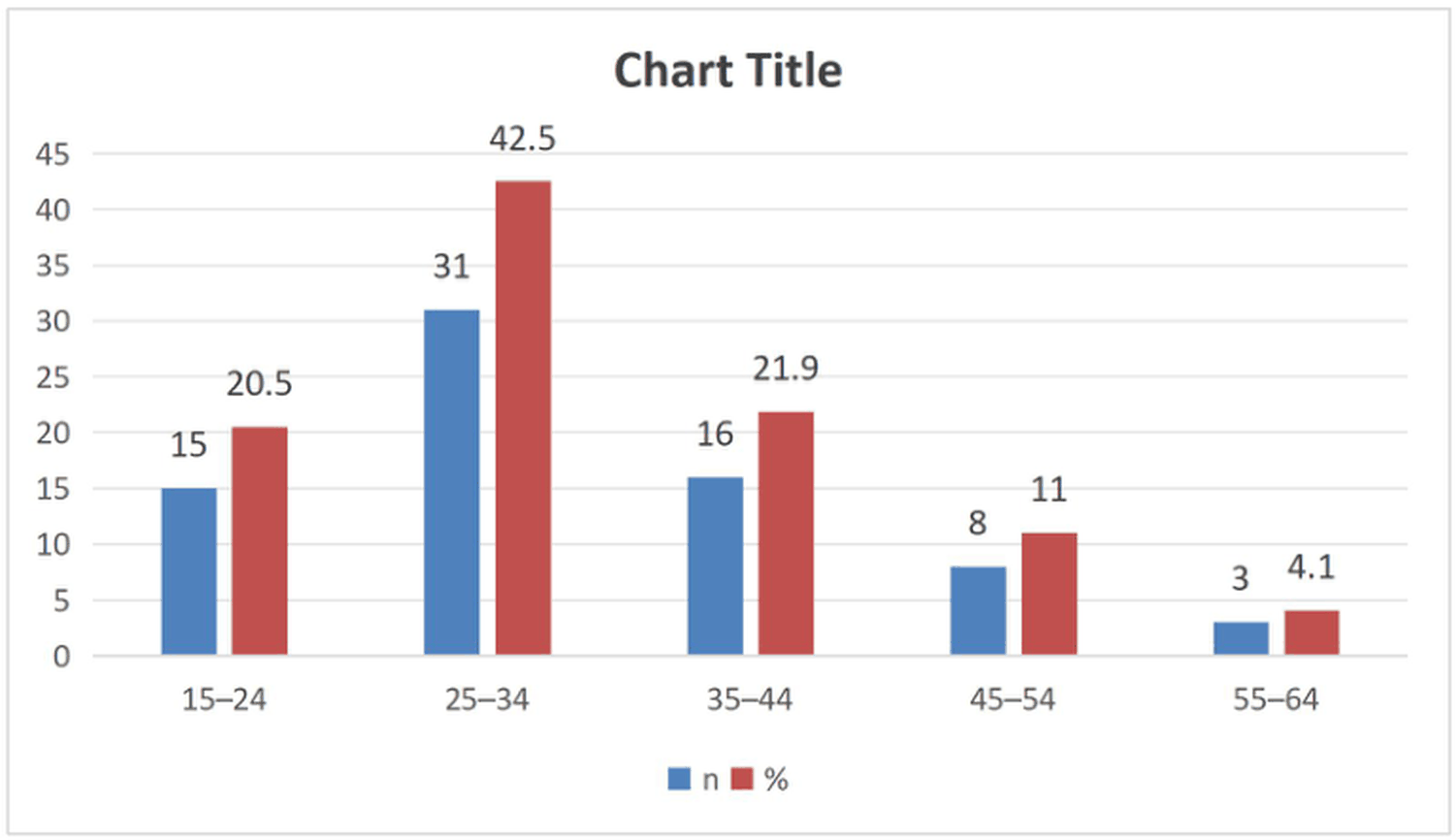 Figure 1: Distribution of age among tuberculosis patients (N = 73).
View Figure 1
Figure 1: Distribution of age among tuberculosis patients (N = 73).
View Figure 1
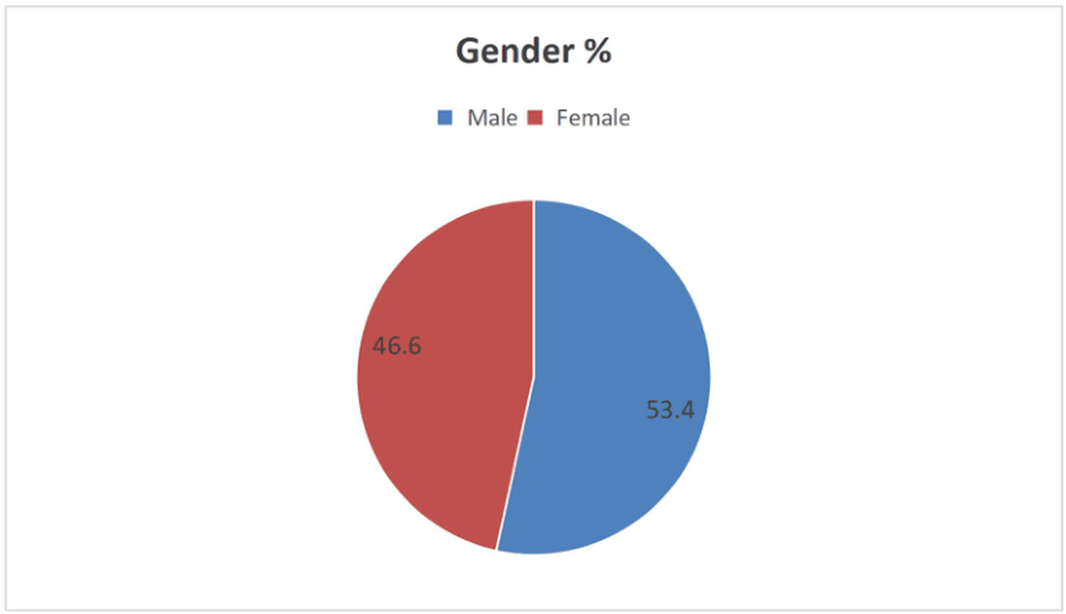 Figure 2: Distribution of gender among tuberculosis patients (N = 73).
View Figure 2
Figure 2: Distribution of gender among tuberculosis patients (N = 73).
View Figure 2
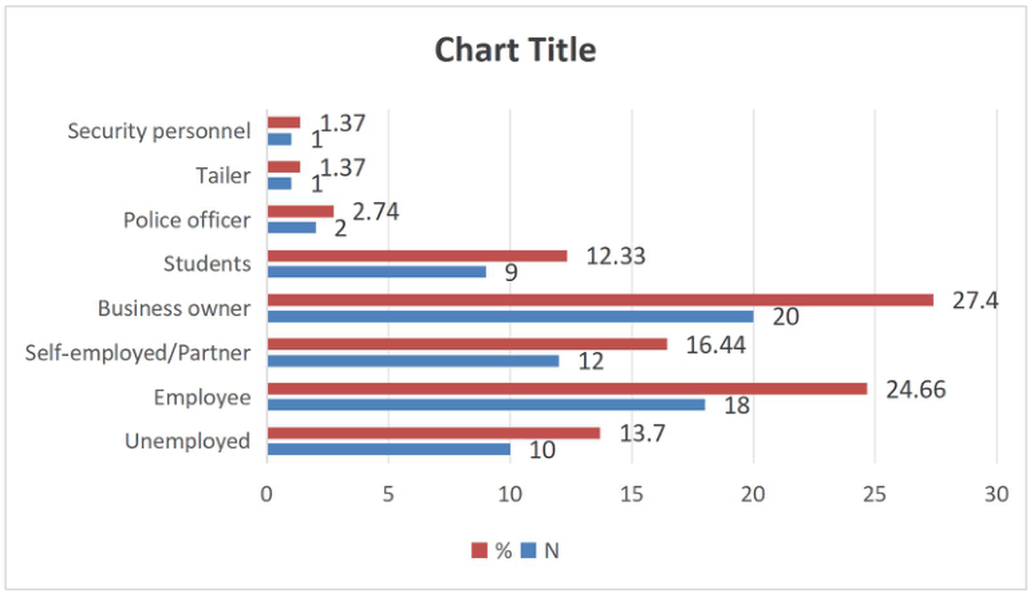 Figure 3: Distribution of occupation among tuberculosis patients (N = 73).
View Figure 3
Figure 3: Distribution of occupation among tuberculosis patients (N = 73).
View Figure 3
Table 2: Participant characteristics (N = 73). View Table 2
It is important to note that none of the patients reported any chronic disease (Figure S4). Only 5 TB patients acknowledged being smokers, while no other patient reported any other negative habit (Figure S5). Almost all of the cohort (94.5%) had no prior history of tuberculosis treatment.
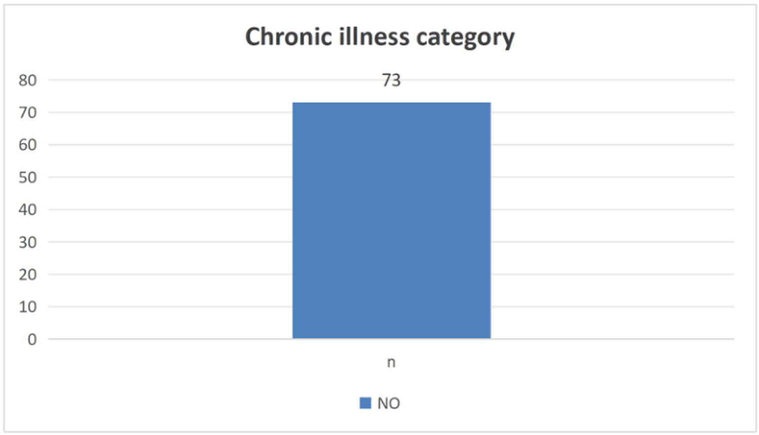 Figure 4: Distribution of chronic illnesses among tuberculosis patients (N = 73).
View Figure 4
Figure 4: Distribution of chronic illnesses among tuberculosis patients (N = 73).
View Figure 4
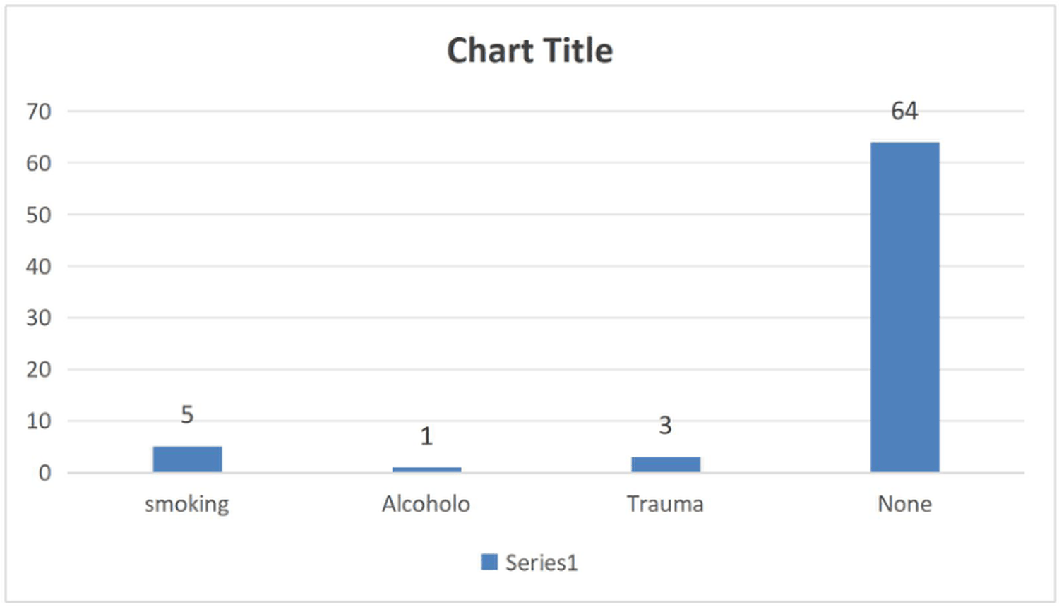 Figure 5: Distribution of bad habit among tuberculosis patients (N = 73).
View Figure 5
Figure 5: Distribution of bad habit among tuberculosis patients (N = 73).
View Figure 5
Furthermore, a vitamin-rich diet was the most common (43.8%) indicator of nutritional status, followed by balanced, diverse meals (38.4%). Table 2 summarizes the participants' characteristics.
Most participants (88.63%) reported no vision changes after commencing ethambutol therapy. The vast majority (84.9%) reported no ocular-toxicity symptoms, while blurred central vision (6.8%) was the leading complaint among those reporting any symptom (Table 3).
Table 3: Self-reported symptoms and ophthalmic findings (N = 73). View Table 3
Visual acuity: Visual acuity within the right eye (OD) for almost all of the participants (94.5%) was normal (6/6). After refraction, visual acuity returned to 6/6 in all participants except one, who remained at 6/24 (Table 3). While left-eye acuity mirrored the right, with 95.9% achieving 6/6 vision. The remaining participants had acuities of 6/12 (1.4%) and 6/18 (2.7%).
Color vision: Color-vision deficits are rare, with only one right eye showing an abnormal result (1.4%). Left-eye color vision was normal in 98.6% of cases, matching right-eye findings.
Pupillary reactions: Normal right-eye pupillary responses dominated (98.6%), with only one participant (1.4%) showing sluggish reaction. Left-eye pupillary reactions were likewise normal in nearly all patients (98.6%). Direct and consensual reactions were normal in 100% of left eyes.
Interocular pressure: The interocular pressure was normal among all of the patients in the study (100%).
Fundoscopy findings: Fundoscopy was unremarkable for 98.3% of participants, with only one participant (1.7%) showing abnormal optic nerve or retinal changes.
Other eye diseases and additional investigations: Most patients (79.5%) had no concurrent eye disease. Each specific pathology appeared in ≤ 3% of the cohort. Dryness was the most common concurrent finding (4.2%), followed by pterygium (1.4%). Only 6.8% of patients required further ocular or systemic investigations beyond the routine assessment. Table 3 summarizes self-reported symptoms and ophthalmic findings.
This cross-sectional study of 73 tuberculosis patients receiving ethambutol in Kassala, Sudan, revealed a notable dissociation between subjective and objective visual findings. While 13.7% of participants reported vision changes and 15.1% described symptoms suggestive of ocular toxicity (e.g., blurred central vision, difficulty reading), objective abnormalities were uncommon: only 1.4% of eyes showed abnormal color vision, 1.4% sluggish pupillary reactions, and 1.7% abnormal fundoscopic findings. Furthermore, nearly all visual acuities were corrected to 6/6 except one patient who remained at 6/24. This pattern suggests that early ethambutol-induced optic neuropathy (EON) may produce subtle perceptual disturbances before manifesting on standard clinical tests, a phenomenon increasingly recognized in the literature.
Our low rate of objectively confirmed EON (1.7% abnormal fundoscopy) aligns with several contemporary cohort studies. For instance, a nationwide cohort study from South Korea by Kim et al. (2024) reported the cumulative incidence of overall optic neuropathy among ethambutol users to be 2.8%, with a 1-year cumulative incidence of 2.1% for optic neuropathy/optic neuritis [6]. Furthermore, another study focusing on subclinical toxicity found that while clinical symptoms were absent, subclinical damage such as retinal nerve fiber layer (RNFL) thickening was present in 13% of eyes, as detected by optical coherence tomography (OCT) [34]. Our finding that subjective symptoms exceeded objective findings by nearly tenfold echoes the work of Taffner et al. [24], who demonstrated that patient-reported visual complaints during ethambutol therapy correlated poorly with formal testing, emphasizing the necessity of routine screening regardless of symptom status [24].
An intriguing and potentially protective observation in our cohort was the absence of any patient with documented chronic diseases such as diabetes mellitus, hypertension, or HIV infection. This is striking given that Sudan's national TB burden includes substantial proportions of HIV co-infection and rising diabetes prevalence [35,36]. Both conditions are well-established risk factors for EON. Diabetic patients on ethambutol have been shown to exhibit significantly increased oxidative stress markers (e.g., malondialdehyde) and decreased antioxidant levels (e.g., superoxide dismutase, catalase, vitamins E and B1), suggesting heightened susceptibility to ethambutol-induced optic neuropathy through impaired mitochondrial reserve [37]. HIV co-infection similarly compromises neural repair capacity. Our cohort's lack of these comorbidities likely explains, at least partially, the low rate of objective EON. Including a more representative sample of Sudanese TB patients, many of whom present with malnutrition and co-morbidities, the toxicity rate might have been considerably higher.
Nutritional status may have further modified our findings. Nearly 44% of participants self-reported a vitamin-rich diet, and another 38% reported diverse meals. This is relevant because ethambutol is a chelator of copper and iron, and its mitochondrial toxicity is exacerbated by deficiencies in B vitamins (particularly B12 and folate) and trace elements. Our cohort's relatively favorable dietary profile may have conferred neuroprotection, potentially explaining why objective toxicity remained low despite a substantial proportion of subjective complaints.
Several actionable implications emerge. First, the poor concordance between subjective symptoms and objective findings means that relying on patient complaints alone will inevitably miss early, reversible EON. Structured screening using Snellen visual acuity, Ishihara color plates, and direct ophthalmoscopy, which are all low-cost, low-technology tools, should be mandatory at baseline, monthly during the ethambutol-intensive phase, and at treatment completion. Second, even a single patient with irreversible visual loss (our one case with residual 6/24 acuity) represents a preventable tragedy; prompt ethambutol discontinuation upon confirmed toxicity can halt progression. Third, given Sudan's high malnutrition prevalence, integrating nutritional assessment and empirical B-vitamin supplementation into TB protocols is a low-cost strategy that warrants formal evaluation.
This study has significant limitations. The cross-sectional design prevents establishing causality or temporal progression. It lacks pre-ethambutol baseline data for most participants. Convenience sampling and a modest sample size (N = 73) limit generalizability and statistical power for subgroup analyses. No advanced diagnostics (OCT, visual evoked potentials, contrast sensitivity, or electroretinography) were available, so subclinical optic nerve damage may have been under-detected. The absence of a control group not receiving ethambutol (ethically challenging) weakens causal attribution. We also did not record the cumulative ethambutol dose or exact treatment duration, both critical determinants of toxicity.
Longitudinal cohort studies with pre-treatment baseline ophthalmic assessments, larger sample sizes, and longer follow-up are urgently needed in Sudan. Future research should evaluate whether routine B-vitamin supplementation reduces EON incidence in malnourished TB populations and should incorporate affordable point-of-care technologies such as smartphone-based fundoscopy. Policy-makers should amend national TB guidelines to mandate regular ophthalmic screening and to establish clear referral pathways when toxicity is suspected.
Ethambutol remains a cornerstone of tuberculosis treatment but carries a well-recognized risk of ocular toxicity, even at standard doses. In this study, although clinically significant objective toxicity was uncommon, a notable minority experienced visual symptom, underscoring the persistent need for routine ophthalmic monitoring. The absence of chronic diseases and the favorable dietary profile in this cohort may have partially protected against more severe toxicity. Given the high prevalence of malnutrition and comorbidities in Sudanese TB patients, enhanced, structured screening protocols are strongly warranted. Early detection and prompt intervention can prevent irreversible vision loss and improve overall TB treatment outcomes. These findings support the systemic integration of ophthalmic care into TB management protocols in Sudan and similar resource-limited settings.
None.
The authors declare no conflict of interest.
The study did not receive any funding.
All authors contribute equally in the preparation of manuscript and approved its final form.
All of the data is present within the manuscript.
Ethical approval was sought from Ethics Review Committee of the Sudan Medical Specialization Board, Ethics Committee at the Research Unit (E.D.C) for approval of the study. The permission to conduct the study was requested from The Ministry of health in Kassala state and then from Administrator of Kassala Teaching Hospital.
Informed consents were obtained from the participants and parental consent was obtained if the participants were younger than 18 years.