
Introduction: Lateral ankle sprains are a common injury that tipically heals quickly with conservative treatment. However, the presence of a severe post-traumatic hematoma can cause significant complications, turning a non-severe injury into a more complex and potentially debilitating process with possible long-term consequences.
Presentation of Case: A young man, with no significant medical history, experienced a rupture of the lateral ankle ligaments. A large post-traumatic hematoma resulted in skin and soft tissue necrosis due to pressure-induced ischemia. This subsequently exposed the underlying structures. The conservative treatment administered was detailed, and surgical intervention was not necessary to resolve the traumatic process.
Discussion: Severe post-traumatic swelling can occur due to vascular ruptures caused by the inversion mechanism and forced plantar flexion leading to serious local complications. The various classification systems for ankle sprains have also been discussed, highlighting their high level of heterogeneity and the absence of an universal classification system to aid in staging.
Conclusion: Ankle sprains are a common and easily treated traumatic injury. However, they can also present associated injuries that can compromise joint function, with potential sequelae.
Ankle, Sprain, Wound, Ligament, Case report
1. Skin necrosis following an ankle sprain is a rarely documented complication, making the identification of warning signs essential.
2. Negative pressure therapy plays a crucial role in the healing of soft tissue after an open injury, potentially allowing patients to avoid surgery.
3. The various classification systems for ankle sprains should be consolidated into one universal system.
1. Skin necrosis following an ankle sprain is a rarely documented complication, making the identification of warning signs essential.
2. Negative pressure therapy plays a crucial role in the healing of soft tissue after an open injury, potentially allowing patients to avoid surgery.
3. The various classification systems for ankle sprains should be consolidated into one universal system.
Lateral ankle sprain is one of the most common musculoskeletal injuries, especially in athletes. Although it is a relatively benign injury, inadequate treatment can lead to residual symptoms and chronic instability [1].
Appropriate management of this injury is essential for successful recovery, and a correct diagnosis based on the precise identification of the different structures involved is essential [2]. High-grade sprains may be associated with bone or chondral injuries. Neurological complications are less common [3], and open ligament injuries without an associated fracture are very rare [4]. Occasionally, a large post-traumatic hematoma can cause necrosis of the skin and underlying soft tissues due to ischemia induced by excessive pressure of the hematoma.
We present a case of a lateral ankle sprain with rare complications unrelated to the treatment method. Additionally, we also review the various classification systems used in the literature to categorize lateral ankle ligament injuries.
This case report has been reported in line with the SCARE Criteria [5].
A 49-year-old male, with no significant medical history and a regular athlete, experienced an indirect trauma to his left ankle while playing basketball. The injury occurred due to an inversion and plantar flexion, resulting in immediate swelling on the anterolateral side of the ankle. He also experienced severe pain and functional impotence, making it impossible to bear weight on the affected ankle while walking. Additionally, there was an area of skin pallor on the anterolateral side of the ankle, indicating hyperpressure from an underlying hematoma. Figure 1 shows the immediate aftermath of the trauma, captured in a photograph taken while the player was still on the basketball court.
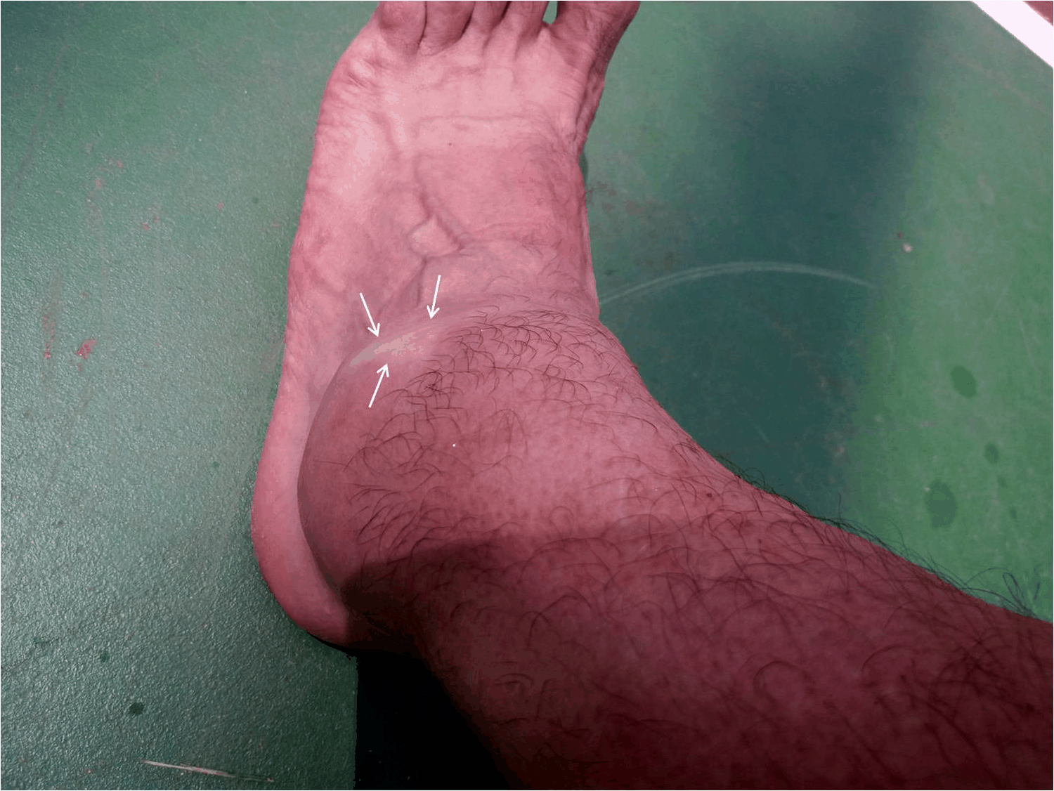 Figure 1: Appearance of the ankle immediately after the trauma, showing significant swelling and an area of skin pallor on the anterolateral aspect of the ankle (white arrows).
View Figure 1
Figure 1: Appearance of the ankle immediately after the trauma, showing significant swelling and an area of skin pallor on the anterolateral aspect of the ankle (white arrows).
View Figure 1
At a major trauma center, an emergency radiological study was conducted to rule out bone lesions. The x-ray confirmed significant soft tissue edema on the anterolateral side of the ankle (Figure 2). Once fractures were ruled out, a severe ankle sprain was confirmed and the patient was diagnosed with a grade III lateral ankle sprain, according to the clinical signs present. The significant initial swelling was considered just one aspect of a serious ankle injury, so no further urgent diagnostic testing was deemed necessary. The ankle was then immobilized with a posterior plaster split, following our center's protocols. Thromboembolic prophylaxis (low molecular weight heparin) was used for 30 days. Additionally, the patient was administered oral anti-inflammatories (dexketoprofen 25 mg, 1 tablet every 8 hours) and analgesics (paracetamol 1 g, 1 tablet every 8 hours) for 7-10 days.
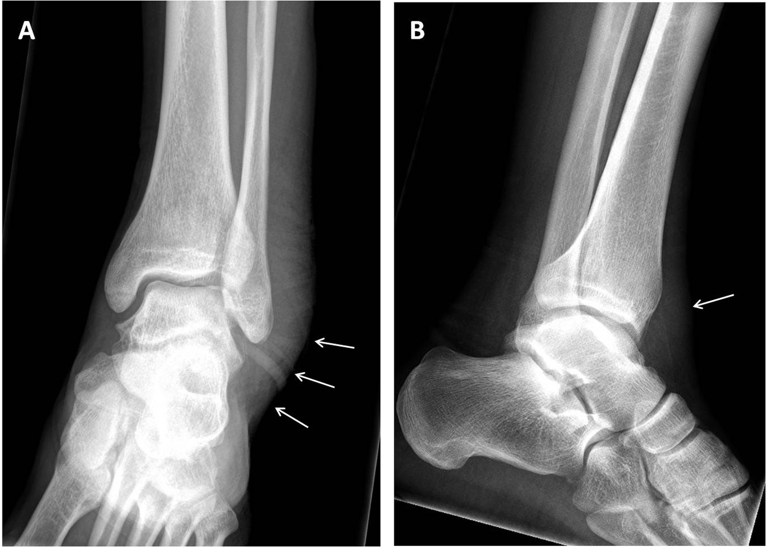 Figure 2: (A): Plain radiographs of the ankle in AP; and (B): lateral projections show lateral and anterior swelling (white arrows), but no signs of bone fractures.
View Figure 2
Figure 2: (A): Plain radiographs of the ankle in AP; and (B): lateral projections show lateral and anterior swelling (white arrows), but no signs of bone fractures.
View Figure 2
Five days after the injury, persistent acute pain that did not respond to prescribed medication prompted a repeat physical examination. The immobilization was removed, revealing early skin lesions on the anterolateral side of the ankle, accompanied with significant swelling in the foot and ankle. However, there was no evidence of acute compartment syndrome requiring further testing. From that point on, treatment focused on the skin condition.
In the following days, examinations confirmed that area of skin previously identified with greater pallor was progressing to cutaneous necrosis (Figure 3). Thirteen days after the injury, the skin eschar was well-defined, requiring debridement and drainage of the underlying organized hematoma. This procedure was performed on an outpatient basis, without the need for anesthesia or hospital admission. The ankle was immobilized with a plaster cast for four weeks.
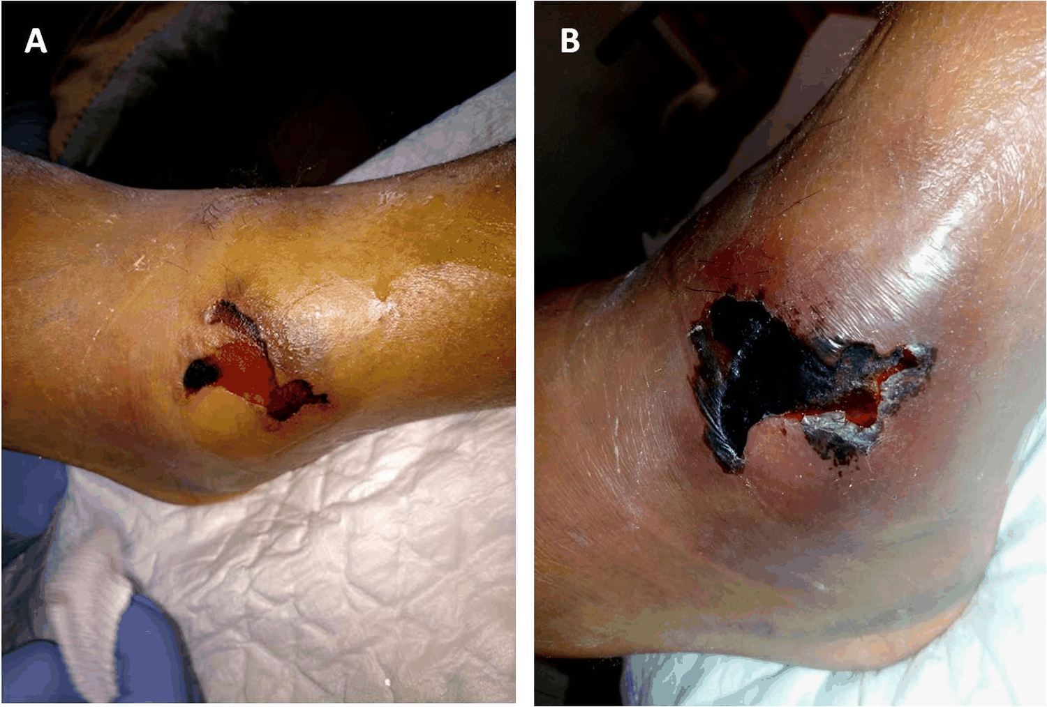 Figure 3: Progression of the skin lesion at (A): 5 days; and (B): 13 days.
View Figure 3
Figure 3: Progression of the skin lesion at (A): 5 days; and (B): 13 days.
View Figure 3
After cleaning, a deep defect was revealed with exposure of the anterolateral capsule-ligamentous plane, converting the injury to an open condition (Figure 4). Outpatient treatment was initiated with PICO negative pressure therapy (NPT) (Smith & Nephew, London, UK) and antibiotic prophylaxis (amoxicillin + clavulanic acid 875 mg, 1 oral tablet every 8 hours) was used for 5 days. This NPT system provided 80 mm Hg of negative pressure to the wound surface, helping to control exudate and promoting secondary intention healing. The skin lesion completely healed after four months, thanks to multiple treatments and replacements of the negative pressure system (Figure 5).
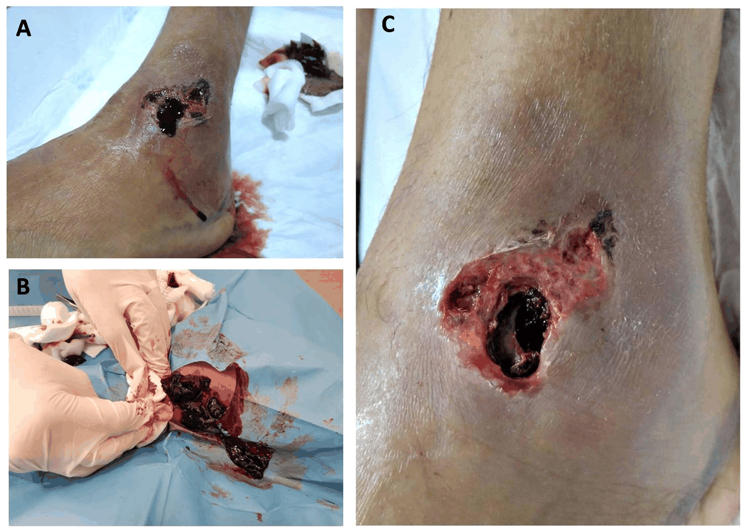 Figure 4: Treatment of the skin lesion: (A): Debridement; (B): Drainage of the organizing hematoma; and (C): Appearance at the end of the procedure.
View Figure 4
Figure 4: Treatment of the skin lesion: (A): Debridement; (B): Drainage of the organizing hematoma; and (C): Appearance at the end of the procedure.
View Figure 4
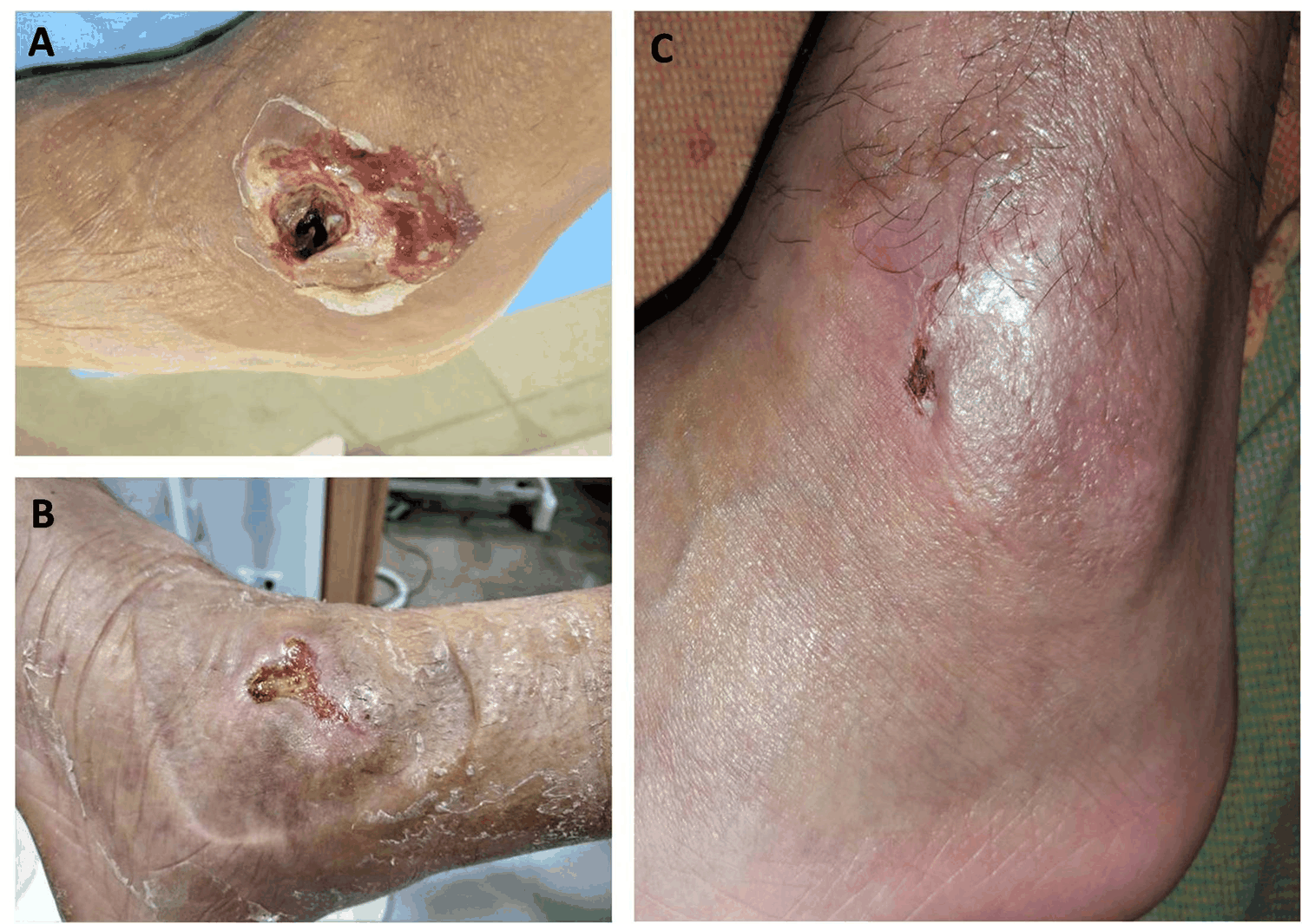 Figure 5: Progression of the skin lesion with negative pressure therapy at (A): 1 month; (B): 2 months; and (C): 4 months.
View Figure 5
Figure 5: Progression of the skin lesion with negative pressure therapy at (A): 1 month; (B): 2 months; and (C): 4 months.
View Figure 5
Concurrently, the patient presented with neuropathic pain starting in the first week, along with an anesthetic area on the dorsum of the forefoot and toes, indicative of a superficial peroneal nerve (SPN) injury. Oral treatment with pregabalin was initiated with a progressive increase in dose. The necrotic area aligned with the subcutaneous path of the SPN on the anterolateral side of the ankle. Electromyography reported neurotmesis of the SPN. We suspect that the nerve injury was likely due to stretching during the initial trauma, involving extreme inversion and plantar flexion of the ankle, compression from a severe post-traumatic hematoma, and subsequent debridement of the area where the SPN typically passes through.
Two months later, a magnetic resonance imaging (MRI) scan was performed. At our center, we are unable to conduct emergency MRIs following an ankle sprain, which presents a significant limitation for the prompt diagnosis of severe injuries. The MRI was performed later, mainly to rule out any lesions in the talar dome that might require secondary surgery and accurately classify the joint injuries. The MRI report revealed tears in the anterior talofibular ligament (ATFL), the calcaneofibular ligament (CFL), and the anterior ankle joint capsule. Bone edema was found along with microfractures in the subchondral region of the medial aspect of the distal tibia and cuboid bone. The posterior talofibular ligament (PTFL) remained intact, and chondral lesions of the talar dome were ruled out (Figure 6-Figure 8).
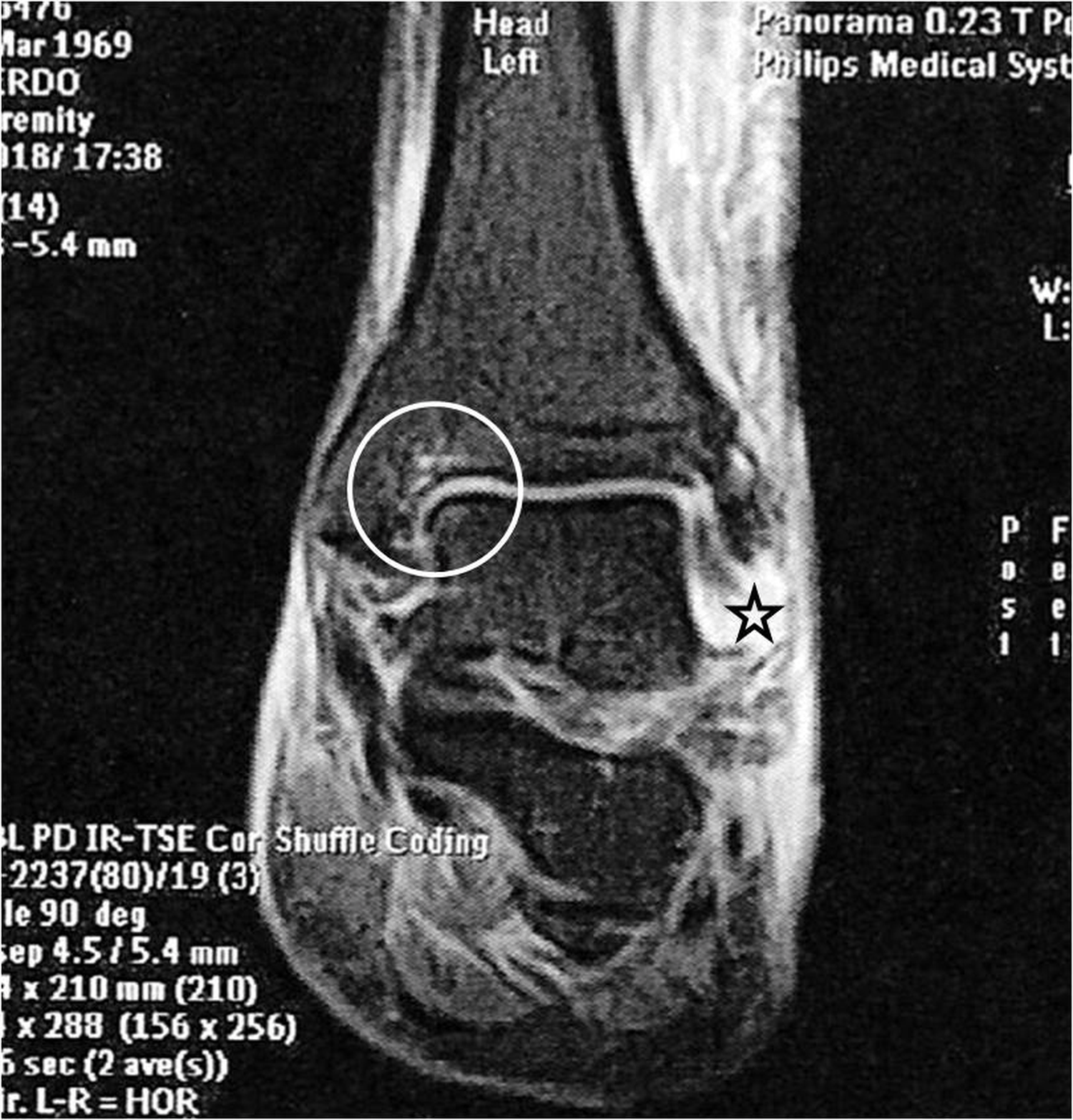 Figure 6: Coronal MRI. Rupture of the anterior talofibular ligament (star) with impaction injuries to the talus and distal tibia in the medial region of the ankle (circle).
View Figure 6
Figure 6: Coronal MRI. Rupture of the anterior talofibular ligament (star) with impaction injuries to the talus and distal tibia in the medial region of the ankle (circle).
View Figure 6
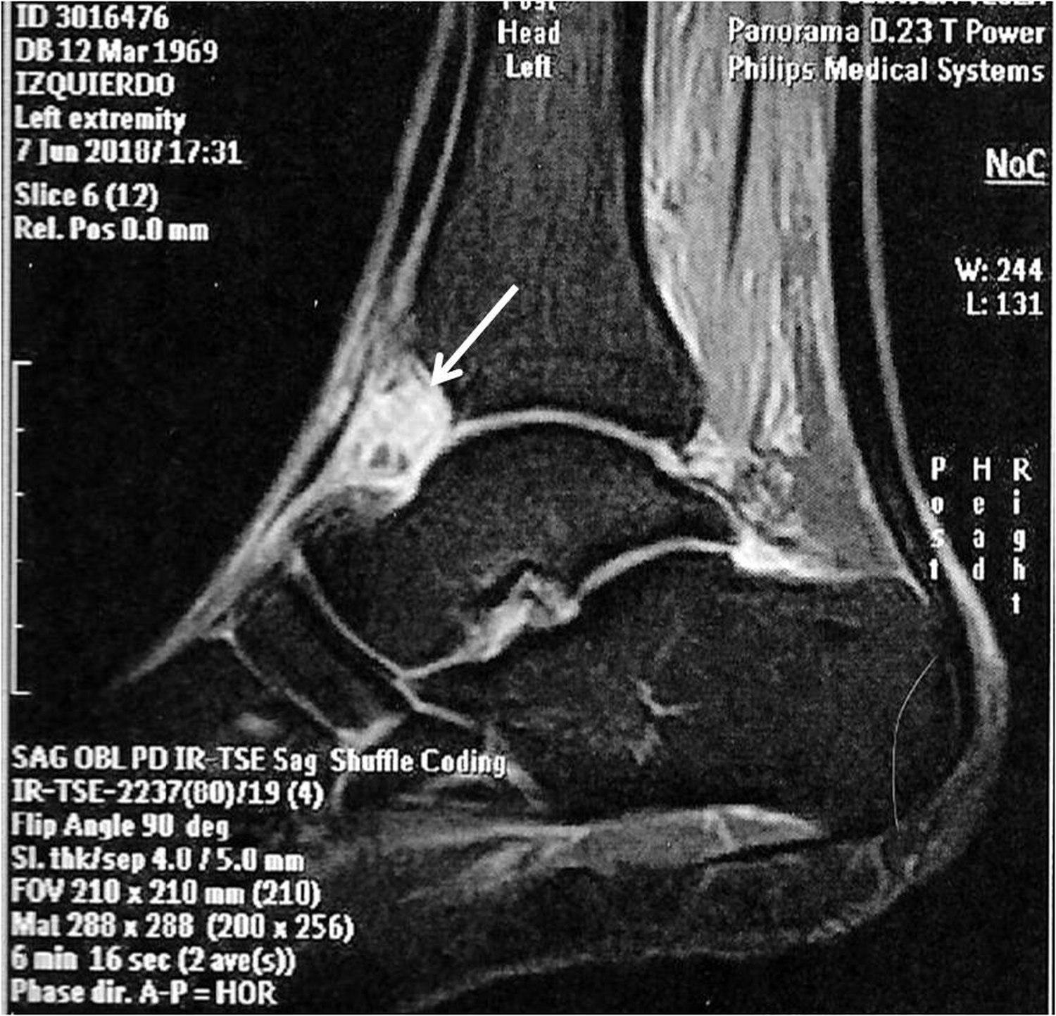 Figure 7: Sagital MRI. Rupture of the anterior capsule (white arrow).
View Figure 7
Figure 7: Sagital MRI. Rupture of the anterior capsule (white arrow).
View Figure 7
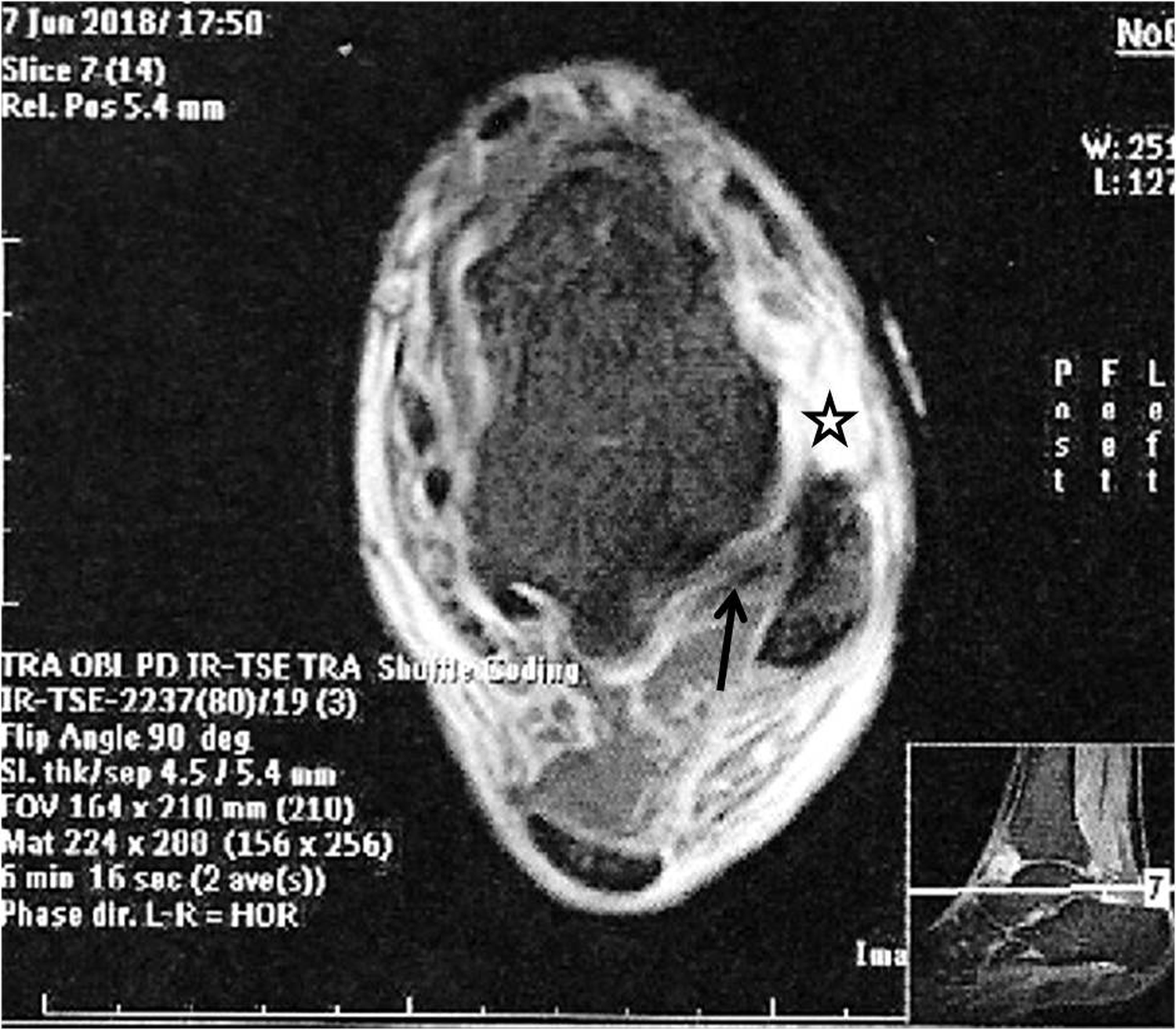 Figure 8: Transversal MRI. Rupture of the anterior talofibular ligament (star) with intact posterior talofibular ligament (black arrow).
View Figure 8
Figure 8: Transversal MRI. Rupture of the anterior talofibular ligament (star) with intact posterior talofibular ligament (black arrow).
View Figure 8
Rehabilitation treatment was intensified after the soft tissue had healed, leading to a full recovery of strength and range of motion. At the 6-month follow-up, the patient demonstrated mechanical and functional stability in the ankle, scoring 100 on the American Orthopedic Foot and Ankle Society (AOFAS) scale and 48 on the Self-Reported Foot and Ankle Score (SEFAS). The most significant medium-term consequence was post-traumatic anesthesia in the foot caused by an injury to the SPN. He was able to return to sports 8 months after the injury. Therefore, a follow-up MRI was not necessary.
Skin necrosis in the development of a lateral ankle sprain is a very rare complication. There are few publications on ankle ligament injuries with cutaneous complications. Souyberand, et al. [4] presented the largest series, with 9 cases of severe open ankle sprain, which they defined as a lateral or medial ligament rupture with an associated transverse tear of the skin over the corresponding malleolus. All cases were due to high-energy trauma, either from traffic accidents or falls from a height. Other publications reported isolated cases of open ligament injuries without dislocation [6,7] or with ankle dislocation [8,9]. In the case presented, the open injury developed gradually during the evolutionary process, rather than acutely. This was due to a severe post-traumatic hematoma causing excessive pressure on the skin of the lateral part of the ankle, as well as the underlying soft tissues. The appearance of post-traumatic hematoma on the anterolateral side of the ankle may be due to the rupture of the vascular branches of the perforating peroneal artery that run between the two bands of the ATFL [10].
Following an ankle sprain, swelling is a common symptom. However, vascular rupture caused by extremely sudden movements can lead to the formation of a significant hematoma. If this hematoma is not reabsorbed, it can become organized, increasing the risk of pressure buildup in an area lacking muscle structures to protect the skin.
The trauma was caused by a severe combined mechanism. Upon reviewing the case, the initial imaging already showed an area of skin damage (a paler area) on the anterolateral aspect of the ankle due to blood pooling from the injury to the vessels surrounding the ruptured ligaments. Consequently, the persistence and disproportionate severity of the symptoms warranted further examination, which revealed the early skin complication. While initial drainage of this area is not included in our emergency department protocols, the procedure would have decompressed the ankle and prevented the subsequent serious complication. Severe ankle edema is generally treated conservatively. We do not consider the aspiration and evacuation of a hematoma following a severe ankle sprain to be a standard procedure. However, as in the case presented, it may be important to pay attention to certain clear signs that indicate the need for more invasive treatments to prevent further complications.
We agree that urgent evacuation of the hematoma relieves tension on the skin. However, due to delayed diagnosis and the subsequent formation of clots, hematomas are very difficult to evacuate by aspiration. Therefore, an incision and evacuation are usually performed, which typically requires urgent hospital admission and general anesthesia. Nevertheless, in this particular case, the procedure could be completed on an outpatient basis. It seems clear that the rarity of the case precluded the application of established solutions based on previous experience, making it impossible to predict the disastrous evolution of the severe ankle ligament injury.
Acute lateral ankle injuries tipically occur when the ankle is inverted and plantar flexed, commonly seen in basketball when the ankle is severely inverted upon landing on another player's foot after a jump. In these cases, the ATFL injury is present in 65-73% of lateral ankle sprains, with involvement of the CFL being associated in 20-40%, while isolated injury of the CFL only occurs in 2%10. Tears of the PTFL are uncommon12. Forced plantar flexion of the ankle may also result in tearing of the anterior capsule, often closely associated with injury of the ATFL [12].
Chondral injuries and bone contusions are common in high-grade sprains, often found in the talar dome [2]. Jennings and Davies [13] discovered that 6.7% of cuboid-related pathologies occur after excessive ankle inversion forces. The SPN is particularly susceptible to stretching from forced ankle inversion with plantar flexion due to its anterolateral position. Mitsiokapa, et al. [3] showed that in high-grade lateral sprains, the SPN is at risk of overstretching, leading to functional impairment, cessation of vascularization, and structural damage to the peripheral nerve with as little as a 15% increase in strain.
Negative pressure wound therapy (NPT) has become an effective and safe tool for treating extensive and complex wounds. NPT systems consist of an open-cell foam sponge and an adhesive dressing, placed over a vacuum pump that generates negative pressure. They are used for tissue defects and chronic wounds as an adjunct to surgical debridement. NPT promotes healing at both the macroscopic and microscopic levels, and in some cases is used instead of more traditional wound treatment techniques, allowing the wound to heal more quickly. This therapy isolates the wound, reducing the possibility of secondary contamination from the environment and minimizing edema. This reduction in edema improves capillary blood flow, increasing the supply of oxygen and nutrients to the wound and combating bacterial growth [14]. In this clinical case, the NPT led to the complete healing of the skin lesion and underlying soft tissues. Although we are unsure about the type of ligamentous and capsular healing that took place, we recognize that the initial immobilization, NPT, and rehabilitation treatment were sufficiently effective, as there was no mechanical instability of the ankle.
Traditionally, lateral ankle sprains have been classified into grades I, II, and III, with grade III representing the most severe injury. Despite the existence of several classification systems, there is no consensus on which one is the most valid and accurate.
The American Medical Society's Standard Nomenclature System [15] classifies injuries based on ligament damage. Grade I is assigned when there is a stretch, grade II for a partial tear, and grade III for a complete tear without specifying the damaged structure. This system is widely used [16], but it mainly focuses on the status of the ATFL, overlooking potential injuries to the CFL and PTFL.
Other authors have used the Anatomical System, which is based on the number of ligaments injured [1,17]. Gaebler, et al. [17] developed a more precise classification system, based on MRI findings, that categorizes injuries according to the affected ligament structure: grade I includes injuries to the ATFL, grade II involves the ATFL and CFL, and grade III is associated with tears in the PTFL. The main drawback of all these classification systems is that, without surgical treatment or high-quality radiographic evidence, there is no objective certainty regarding the type of ligament injury.
Some authors have based their classification on clinical signs and the degree of instability [18]. For instance, a grade I or mild injury implies mild edema, minimal or no loss of mobility, and no mechanical instability of the joint. A grade II or moderate injury shows more intense pain, lateral swelling with minimal reduction in joint movement, and mild to moderate joint instability. A grade III or severe injury presents marked swelling and edema, significant pain on palpation, reduced range of motion, and obvious instability. The main limitation of this scale is the subjectivity due to variability in clinical interpretation. Other authors [19] have designed different systems considering the ability to bear weight, although they have not had a significant impact on the literature.
Malliaropoulos, et al. [20] developed a more comprehensive classification system based on loss of function, ligament laxity, presence of hematomas, punctate tenderness, loss of movement, and magnitude of edema. The authors discovered that this system could predict the return to sports activities based on the severity of the injury. This classification system has been referenced in other publications [2], although it is more intricate and constrained by the subjective interpretation of physical signs. Additionally, performing anterior drawer and forced inversion maneuvers can be challenging due to acute joint inflammation, which reduces their diagnostic accuracy. Research has indicated that delaying the physical examination (4 to 5 days after trauma) can result in a more accurate diagnosis compared to conducting the examination within 48 hours after the trauma [21].
Therefore, depending on the classification used, the present clinical case could be categorized in various ways.
Although these statements should be treated with caution, as is inherent to case reports, an accurate and comprehensive diagnosis is crucial for lateral ankle ligament injuries. Therapeutic strategies and recovery time are directly related to the severity of the clinical presentation and any associated injuries. These factors can impact the functional prognosis and, as seen in the case presented, potentially delay full recovery.
Written informed consent was obtained from the patient for publication and any accompanying images. A copy of the written consent is available for review by the Editor-in-Chief of this journal on request.
This case report was conducted in accordance with institutional policies. Ethical approval was not required as per the guidelines for single patient case reports.
This study does not qualify as a First in Man study and thus does not require registration under that category.
This research did not receive any specific grant from funding agencies in the public, commercial, or non-profit sectors.
The authors declare that they have no conflicts of interest related to this work.
None.
This research did not receive any specific grant from funding agencies in the public, commercial, or not-for-profit sectors.
This work has not been previously published nor is it under consideration for publication elsewhere. Its publication is approved by all authors, as well as tacitly or explicitly by the responsible authorities where the work was conducted. If accepted, it will not be published elsewhere in the same form, in English or any other language, including electronic formats, without the written consent of the copyright holder.
☒ The authors declare that they have no known competing financial interests or personal relationships that could have appeared to influence the work reported in this paper.
☐ The authors declare the following financial interests/personal relationships which may be considered as potential competing interests: