
Tracheostomy is a common procedure used in patients unable to wean from mechanical ventilation. Complications are rare but dangerous, ranging from bleeding and infection to equipment-related failures. Here, we report a case of secondary airway obstruction caused by tracheostomy cuff herniation over the end of the tracheostomy tube in a polytrauma patient. This report highlights the importance of prompt recognition and management of equipment-related tracheostomy complications and proposes a diagnostic framework for clinicians encountering similar challenges.
Tracheostomy is a common surgical procedure used to progress the care of orotracheally intubated patients unable to liberate from the ventilator. Approximately 100,000 tracheostomies are performed annually in the United States [1]. A frequent indication for tracheostomy in trauma patients is inability to wean from mechanical ventilation [2], with tracheostomy performed on a semi-elective basis. This is generally done for one or both of two reasons: neurologic [3] and/or respiratory [4] impairment causing prolonged requirement for endotracheal intubation. Tracheostomy can decrease rates of pneumonia [5,6] reduce ventilator days [7], assist with work of breathing, and improve patient mobilization [8]. Indications for urgent tracheostomy in the trauma patient population include refractory airway obstruction, penetrating laryngeal trauma, post-cricothyrotomy management, severe facial trauma, and upper airway obstruction, which is the most frequent indication in the emergent setting [9]. Complications of tracheostomy [10] include bleeding, tracheal stenosis, infection, mechanical failure, and fistulization of the trachea to the esophagus or innominate artery, with an overall complication rate of about 3% [11]. Mortality rates directly attributable to tracheostomy placement are low, approximately 0.17% [12]. Some causes of mortality include tracheoinnominate artery fistulas [13,14], which can have a 65% mortality [15], and tracheostomy dislodgement that occurs in 0.8% of cases within one week postoperatively and can result in mortality if not promptly recognized and treated [16]. While surgical techniques to reduce anatomical complications have been well documented [17-19], there remains limited literature addressing equipment-related failures. Rescue from acute mechanical failure requires timely identification and correction. This report presents a case of tracheostomy cuff herniation leading to acute airway compromise and discusses strategies for rapid evaluation and management.
A 35-year-old male was transported via ambulance to our Level I trauma center, following a high-speed motor vehicle collision. He was a restrained backseat passenger with a history of intermittent cocaine use. Upon arrival, a computed tomography (CT) scan revealed extensive injuries including: left 1st-5th rib fractures with dislocation of the 1st rib at the sternocostal junction; a large pneumothorax; a 4-cm distal abdominal aortic pseudoaneurysm extending into the common iliac arteries; and a complex abdominal wall disruption with herniation of the small bowel into the abdominopelvic subcutaneous tissue. Massive transfusion protocol was initiated for hypotension, and the patient was taken emergently to the operating room for exploratory laparotomy. The aortoiliac pseudoaneurysm was repaired using an interposition graft. An inferior vena cava (IVC) injury was repaired, and resection of multiple destructive bowel injuries were performed. The abdomen was temporarily closed at the initial operation. The patient's course was subsequently complicated by abdominal wall necrotizing soft tissue infection and anastomotic revisions, requiring a total of 8 operations over the course of 2 weeks, prior to tracheostomy placement.
The patient's severe chest trauma, including rib fractures and pulmonary contusions, body habitus (body mass index (BMI): 41.4), and deconditioning due to prolonged critical illness complicated his respiratory recovery. In the setting of prolonged intubation in a patient with a very complicated surgical course, an open tracheostomy was planned [20-22].
On hospital day 16, the patient underwent elective open tracheostomy placement with a Shiley 6 XLT tube in the operating room. The case was largely uncomplicated and following placement of the tracheostomy under direct visualization, the cuff was inflated with 15 mL of air. However, the patient then rapidly desaturated, with negligible tidal volumes delivered by mechanical ventilation. Bag-valve ventilation was immediately initiated through the tracheostomy and was notable for palpable resistance to ventilation. Simultaneous maneuvers were immediately performed to assess and treat possible airway obstruction. Preparation to reintubate orotracheally proceeded at the same time as maneuvers to confirm tracheostomy position and relieve obstruction. Direct laryngoscopy demonstrated profound edema of the upper airway. Bronchoscopy was performed through the tracheostomy, confirming correct position of the tracheostomy in the trachea. Bronchoscopy also identified herniation of the cuff over the end of the tracheostomy tube, causing airway obstruction. Upon deflation of the cuff, oxygen saturation, tidal volume, and end-tidal CO2 normalized. The cuff was then partially reinflated and secured, resulting in stable ventilation parameters and normal vital signs.
The remainder of the patient's hospital course included care of his complex injuries, but he was ultimately weaned from mechanical ventilation. The tracheostomy was replaced with an uncuffed tube 60 days after tracheostomy placement, and subsequently decannulated 65 days after initial tracheostomy placement without issue. He was discharged home with a hospital stay of 100 days with outpatient follow-up and rehabilitation.
This case illustrates a rare but critical complication of tracheostomy: mechanical failure due to partial cuff herniation over the end of the tracheostomy tube leading to airway occlusion. Intraoperative bronchoscopy examination revealed that the tracheostomy tube was appropriately positioned but that the cuff had inflated asymmetrically and protruded around the tracheostomy tube, obstructing the airway lumen (Figure 1a and 1b). This herniation was likely due to airway edema in the setting of prolonged intubation, multiple reintubations, and volume overload in the setting of severe ongoing inflammation from multiple critical injuries and severe secondary infection. It is less likely that the cuff herniation was due to overinflation, as the 15 mL of air was within expected volume for inflation. The external pressure from an inflamed airway caused the balloon to protrude over the tube, despite normal volume in the balloon, consequently compromising airway patency. Resolution was achieved by reducing cuff inflation to a volume that maintained tube position without compromising airway patency.
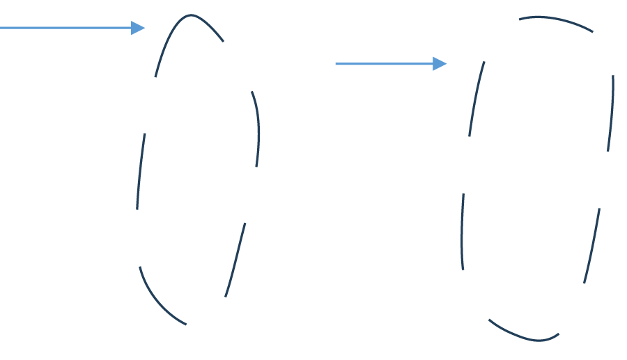 Figure 1: (a) Model of cuff overinflation using a clear plastic syringe to model the trachea. Lateral view of cuff herniating during partial herniation; (b) Superior view of cuff herniating during partial herniation.
View Figure 1
Figure 1: (a) Model of cuff overinflation using a clear plastic syringe to model the trachea. Lateral view of cuff herniating during partial herniation; (b) Superior view of cuff herniating during partial herniation.
View Figure 1
Cuff inflation targets a pressure of approximately 20-30 mmHg to maintain position and ventilatory seal [23]. However, this measurement is generally done in the ICU and not commonly performed intraoperatively [24]. Immediate post-tracheostomy respiratory issues require the operative team to maintain a heightened clinical awareness of potential complications. Ventilatory or oxygenation issues require an assumption that there is surgical or device technical failure and mandate the ability by the surgical team to troubleshoot unexpected equipment-related airway obstructions promptly.
Numerous surgical approaches have been described to assist with management of anatomical complications that can arise during tracheostomy placement. Bleeding is the most common complication and can be managed with vasoconstrictive anesthetic such as lidocaine with epinephrine and local hemostatic strategies such as gauze and direct pressure [19]. Pneumothorax and subcutaneous emphysema are other life threatening complications that can occur in the acute postoperative setting in approximately 1% of cases [25], however these are generally associated with percutaneous placement of tracheostomies rather than open placement [26]. These complications are mitigated by operator experience, limiting comorbidities such as anatomic spinal abnormalities and obesity, and use of smaller bronchoscopes with lower ventilation pressures and tidal volumes. Tracheal wall perforation is another surgical complication, often caused by guidewire instability [27] in percutaneous placement, and can be difficult to manage retroactively. Management is complex and can include either conservative or surgical measures.
There also exists rare complications related to cuff overinflation which can be detrimental if not noticed and appropriately treated. Examples of this include pressure necrosis, ulcers, and fistulas from high intratracheal pressure which can be fatal if not rapidly recognized and managed [27-29]. Long standing ischemia from cuff overinflation can also lead to tracheal stenosis, which can be surgically challenging to manage due to excessive scar tissue [30]. Cuff overinflation can be mitigated by frequent checks of cuff pressure, with maintenance at approximately 20-30 mmHg [24]. Mechanical failures are also possible such as accidental decannulation [31,32] (AD), with some research suggesting occurrence in around 15% of placements. Tube obstruction occurs in approximately 6% of tracheostomies [33]. The likelihood of these failures occurring can be reduced by suturing the tube in place to reduce AD, and humification and suctioning to lower chances of obstruction.
It is important to have a systematic approach to diagnosing and rectifying acute mechanical failure of a tracheostomy. Our diagnostic approach for this case included:
Establishment of a functional, definitive airway is the most important consideration during intraoperative suspected mechanical failure of the tracheostomy. The airway team (anesthesia team in the operating room or the head-of-bed team in bedside procedures) must remain ready to emergently reintubate the patient in the case that airway compromise cannot be alleviated. This is not possible in patients with severe laryngeal edema or laryngeal obstruction, such as those status post laryngectomies.
Trans-tracheostomy bronchoscopy allows for direct visualization and is one of the most effective diagnostic tools to evaluate appropriate placement and patency of the tracheostomy.
Monitoring of cuff leak can be used in placement to determine if cuff is underinflated or damaged. Notably, a persistent cuff leak can be due to cuff herniation through the vocal cords [34], which can lead to permanent damage if not identified and corrected.
Adjust inflation of the tracheostomy cuff based on ventilatory feedback and patient response. Manometry can also be used to guide management for tubing sizing [35].
Tracheostomy remains a critical intervention in patients who cannot liberate from endotracheal intubation. However, equipment-related complications such as cuff herniation, while rare, can result in life-threatening airway obstruction. Clinicians must be prepared to diagnose and resolve such complications rapidly.
I would like to express my sincere thanks to my advisor, Dr. Demetriades, for his guidance and feedback throughout this research project. I am also grateful to Dr. Hodges and Dr. Siletz for their insightful feedback and discussions. Finally, thank you to Monica Wong for being an incredible mentor and support throughout the research process. Thank you to Los Angeles Medical Center and Keck School of Medicine for their support in medical research.
This research did not receive any specific grant form funding agenices in the public, commercial, or not-for-profit sectors.