
Primary ovarian insufficiency (POI) can be a devastating diagnosis with significant psychological stress and life-long health problems including cardiovascular disease, osteoporosis, and diminished life expectancy. We present a case of a 26-year-old adult female (XX) with distal deletion of chromosome 9p who presented with secondary amenorrhea for five years. She had typical dysmorphic features of 9p deletion syndrome. She required a G-tube for nutritional support and was non communicative. Her BMI was 16 and initial consideration was hypothalamic amenorrhea. Her FSH was 25mIU/mL, estradiol was 6 pg/mL, total testosterone was 11 ng/dL, and prolactin was 11 ng/mL consistent with primary ovarian insufficiency (POI). This is now the second reported case of an adult XX individual and distal 9p deletion to present with primary ovarian insufficiency. The DMRT1 gene loci is located on the distal portion of chromosome 9p. This is further evidence that abnormalities (hemizygosity) of DMRT1 may be associated with POI and possibly causative. Adult patients with distal 9p deletion should be screened for POI and, when present, hormone replacement should be considered.
Primary ovarian insufficiency, 9p distal deletion syndrome, Hormone replacement therapy
The American Society for Reproductive Medicine (ASRM) and European Society of Human Reproduction and Embryology (ESHRE) define primary ovarian insufficiency (POI) as loss of ovarian activity before age of 40 years. It is characterized by amenorrhea or irregular menstrual cycles with elevated gonadotropins and low estradiol [1]. POI is a devastating diagnosis with long-term psychological and health consequences [1]. Etiology can be iatrogenic (gonadotoxic chemotherapy, radiation, or surgical oophorectomy), genetic (Turner’s syndrome), but is commonly idiopathic [1]. We report a case of an adult female with 9p deletion syndrome who presented with secondary amenorrhea and findings consistent with POI. We discuss the potential etiology and the need to consider hormone replacement in all patients with POI.
The patient was a 26-year-old Gravida 0 who presented to her local gynecologist with secondary amenorrhea for 5 years. She was previously diagnosed with 9p deletion and had typical dysmorphic features including trigonocephaly and facial dysmorphism. She was noncommunicative and had intellectual disability. She had a G-tube in place for nutritional support. She had previously undergone splenectomy for splenomegaly. CT scan of abdomen/pelvis one year prior to presentation demonstrated unremarkable pelvic imaging with a normal uterus noted. Her BMI was 16. Initial differential included hypothalamic amenorrhea with her low BMI. Her karyotype was 46 XX, del (9) (p22) (Figure 1). More detailed genetic analysis was unavailable. Her FSH was 25miu/mL, estradiol was 6 pg/mL, total testosterone was 11 ng/dL, prolactin was 11 ng/mL consistent with primary ovarian insufficiency (POI). AMH and LH levels were not obtained. Hormone replacement therapy was discussed with the patient’s parent for cardiovascular and bone health. A regimen of transdermal estrogen along with continuous progestin was offered to minimize vaginal bleeding (as opposed to cyclic progestin).
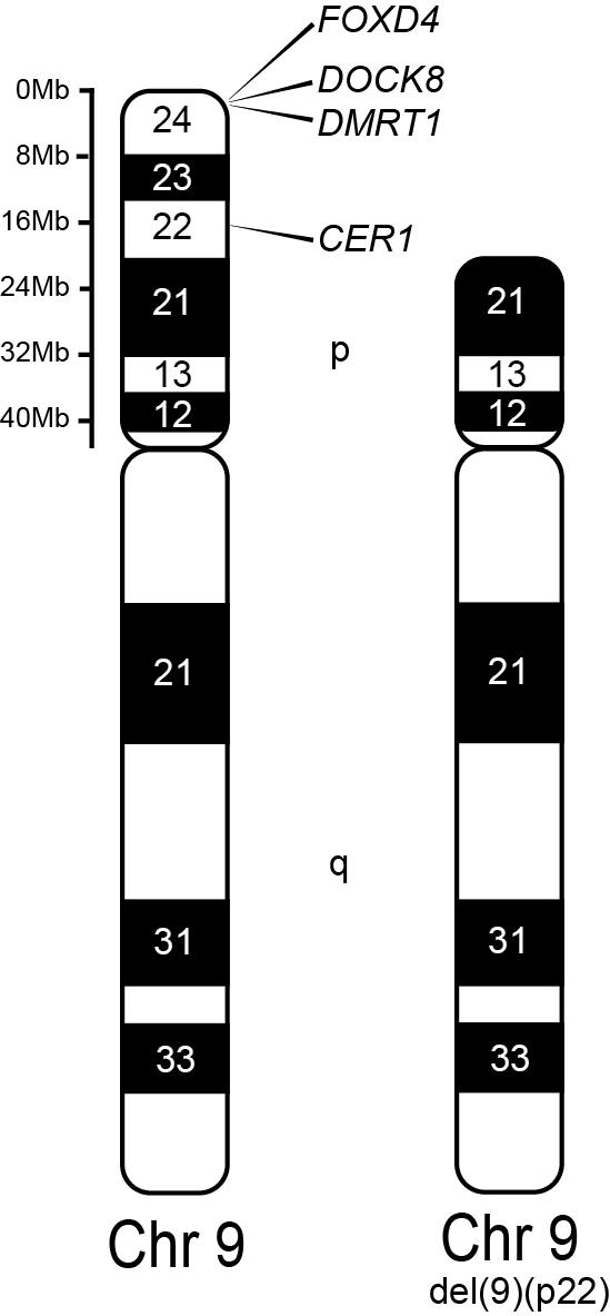 Figure 1: Visual gene map of chromosome 9 highlighting the location of DRMT1 gene and distal deletion present in our patient.
View Figure 1
Figure 1: Visual gene map of chromosome 9 highlighting the location of DRMT1 gene and distal deletion present in our patient.
View Figure 1
9p deletion is a well-known syndrome especially in the pediatric and medical genetic realm. The Online Mendelian Inheritance in Man database describes two different phenotypes for patients with deletions in the short arm of chromosome 9. The “9p deletion syndrome” (OMIM# 158170) is characterized by trigonocephaly, moderate to severe intellectual disability, and facial dysmorphism. The “male to female sex reversal” (OMIM# 154230) presents as abnormal development of the external genitalia in XY individuals with sex reversal [2]. These may be present individually or in combination. The 9p deletion syndrome occurred with deletion of 9p22.2-p23 [3]. Minor to complete gonadal dysgenesis can result from monosomy of 9p24.3 [4] with hemizygosity for DMRT1 (doublesex and mab-3 related transcription factor) highly suggestive for gonadal phenotype of XY individuals [5].
Hauge et al. mapped the breakpoints of 10 patients with 9p deletions and correlated their results to phenotypic features of 9p deletions. They identified multiple genes that contribute to typical features of 9p deletion syndrome, including CER1, which may contribute to trigonocephaly, FOXD4, which may play a role in speech and language or neurological development, and DOCK8, which is a potential guanine nucleotide exchange factor, whose deletion may play a role in seizures. Their findings also indicated that the deletion of DMRT genes alone does not necessarily lead to sex reversal in 46XY individuals [6].
There is extensive research regarding the role of DMRT1 in male gonadal differentiation, but little is known about the role of DMRT1 in female gonadal development. Most reports on 9p deletions are on fetuses and children and the role of DMRT1 in female gonadal function is unknown. Bartels et al. reported on a 31-year-old infertile woman presenting with primary ovarian insufficiency, mild dysmorphic features, and a history of mild developmental delay [7]. Her karyotype was 46XX, del(9)(p23-24). We present a case of a 26-year-old adult female with secondary amenorrhea for five years and hormone testing consistent with POI. Her karyotype was 46XX, del(9)(p22). She had features consistent with “9p deletion syndrome.” To our knowledge, these are the only two cases of adult XX patients hemizygous for the DMRT1 that presented with POI. Most reports on XX individuals with 9p deletion have been in fetuses and children with variable ovarian phenotypes ranging from primary ovarian dysfunction to menses [8,9]. Taken together, these data would suggest that children with hemizygosity of DMRT1 who have gonadal function into adulthood may be at risk for POI and implicates abnormalities or absence of one copy of the DMRT1 gene as a potential genetic cause of POI, but additional studies are required to determine the precise role of DMRT1 in female gonadal function.
A cohort study of 1,858 women born between 1928 and 1932 reported the prevalence of POI as 1% [10]. Studies from North America and Sweden reported the incidence of POI to be between 1.1% and 1.8% [11,12]. However, more recent meta-analyses of the global prevalence of POI reported a rate of 3.5-3.7%, and it appears to be increasing [13,14]. We suspect the 9p deletion with hemizygosity of the DMRT1 gene may be the etiology of POI in our patient. Although chromosomal deletions increase with advancing reproductive age [15,16], we don’t believe this explains the increasing prevalence of POI globally. However, it may be related to advancing reproductive age by another etiology. Increased maternal age has been shown to have a detrimental effect on oocyte mitochondrial health and reproductive outcomes [17]. As offspring get their mitochondrial pool from the oocyte (which have the largest number of mitochondria of any cell [18]), children of older mothers will, in general, have less healthy mitochondria than children of younger mothers. This can also lead to a mutigenerational, compounding effect (i.e., the children of older mothers whose mothers were older could further diminish mitochondrial health). As mitochondrial health is related to reproductive aging, increasing this age for multiple generations may have a compounding effect and contribute to the apparent increase in prevalence of POI.
Regardless of the reason for increasing prevalence of POI, it is a devastating diagnosis with long-term psychological and health consequences for which hormone replacement therapy (HRT) should be considered. [1,19,20]. Women with POI are at higher risk for bothersome menopausal symptoms, reduced bone density and fractures, shortened lifespan primarily due to cardiovascular disease, depression, anxiety, cognitive decline, and dry eye syndrome [1,19,20]. HRT has clear benefits to bone health, cardiovascular protection, menopausal symptoms, and cognitive function [1,19,20]. Physiological transdermal administration may be preferred due to lower thromboembolic risk and potentially better cardiovascular protection [19]. Androgens may be helpful for hypoactive sexual disorder [1]. It would be difficult to assess the psychological impact of HRT in our patient. However, the overall physical health benefits necessitate its consideration in all patients with POI.
We chose to offer a regimen of transdermal estrogen with continuous progestin to our patient. Transdermal estrogen has the benefit of ease of use in a patient with disabilities along with better thromboembolic and cardiovascular profile compared to oral estrogen. Continuous progestins were chosen to minimize risk of vaginal bleeding which can be distressing to patients with disabilities as well as their caregivers.
Both ARSM and ESHRE recommend genetic testing in all patients who present with signs and symptoms of POI that are not iatrogenic [1]. These include karyotype and testing for FMR1 premutation [1]. Karyotype analysis will detect significant 9p deletion as in our patient. As gonadal dysfunction can vary in XX individuals with 9p deletion [7-9], we would recommend screening adult patients with this syndrome with menstrual history, gonadotropin, and estrogen levels.
9p deletion syndrome is a well-known condition especially in the pediatric and medical genetics fields. It can present a unique circumstance for the gynecologist. Hemizygosity of the DMRT1 gene in XX adult females appears to put these individuals at risk for POI which can reduce both quality of life and life expectancy. All women with the presentation of POI should have a genetic workup including karyotype and assessment of the FMR1 premutation. Further studies will be needed to determine the precise role, if any, of DMRT1 mutations in the development of POI. HRT can improve both quantity and quality of life and should be considered for all patients with POI.
No Artificial Intelligence Generated Content (AIGC) tools were used for developing any portion of this manuscript.
None of the authors have any financial disclosures.