
Balamuthia mandrillaris infection and chronic subdural hematoma are distinct medical conditions that can intersect, particularly in immunocompromised patients or those with central nervous system complications.
Risk factors for chronic subdural hematoma include advanced age, the use of anticoagulants, alcoholism, and mild head trauma. Symptoms may consist of headache, confusion, limb weakness, seizures, and altered consciousness. While there is no direct connection between Balamuthia mandrillaris and chronic subdural hematoma, these conditions can sometimes occur simultaneously, and their neurological symptoms may obscure each other. The present report highlights a patient with chronic subdural hematoma with neurological signs, revealing an incidental histopathological diagnosis of Balamuthia mandrillaris infestation.
İntracranial, Parasitic disease, Treatment, Balamuthia mandrillaris
Chronic subdural hematoma (CSDH) has an incidence rate of 1.7 to 20.6 per 100,000 people per year. Balamuthia infections have been reported in over 200 cases worldwide and are most common in immunocompromised individuals and the elderly. Balamuthia mandrillaris (BM) was first identified in 1986 in a mandrill that died of meningoencephalitis, and human infections were described in 1991 [1,2,3]. Although rare, it is usually fatal and typically leads to granulomatous amoebic encephalitis (GAE). The amoeba has two stages in its life cycle: trophozoite and cyst. The trophozoite is 12-60 μm in diameter. The cyst is a round cell measuring 6 to 30 μm [3]. In this report, we present a rare case initially diagnosed as CSDH but later confirmed as BM by histopathological examination. This is the second intracranial Balamuthia mandrillaris case reported in Türkiye following the initial detection in 2009 [4].
A 71-year-old male patient, a retired naval officer, was admitted to the hospital with complaints of weakness in his right arm and leg, difficulty speaking and walking, numbness, and loss of balance. His medical history included alcohol use, balance disorders, and related head trauma. A neurological examination revealed a Glasgow Coma score (GCS) of 12, right hemiparesis, dysarthria, numbness, and an ataxic gait. Laboratory results showed Hb 13 g/dl, hematocrit 38.3%, erythrocytes 4.29 M/µl, leukocytes 6.89 b/µl, no parasites or eggs found in stool, and cystic hydatid hemagglutination was negative. Magnetic resonance imaging (MRI) showed a 3.8 cm diameter subdural hemorrhage in the left frontal and parietal regions, adjacent to the temporal and occipital regions. The hemorrhage contained both thin and thick septa, resulting in midline displacement and mass effect. In the case diagnosed with CSDH, craniotomy was performed in the left parietotemporal region instead of hole drainage, which carries a 20% risk of recurrence (Figure 1a, Figure 1b and Figure 1c). Upon operation, the membranes covering both the calvarial and cortical surfaces were found to be 1-2 mm thick and easily detachable. These membranes were then partially excised. Additionally, a subdural hematoma measuring 5 cm thick, which contained both liquid and solid components along with septal remnants, was aspirated. Microscopic examination revealed hemorrhagic and necrotic tissues, along with areas of eosinophilic meningitis rich in polymorphonuclear leukocytes (PNLs), which contained dense eosinophils and mononuclear cells.
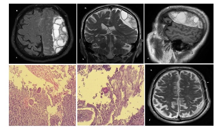 Figure 1: The Axial and coronal T2-weighted (a, b) Sagittal T1-weighted MR images (c) Microscopic examination; scattered amoeba cells (d,e) Postoperative MRI scans at 1.5 months and 2 years (f).
View Figure 1
Figure 1: The Axial and coronal T2-weighted (a, b) Sagittal T1-weighted MR images (c) Microscopic examination; scattered amoeba cells (d,e) Postoperative MRI scans at 1.5 months and 2 years (f).
View Figure 1
Scattered amoebic cells, ranging in diameter from 10 to 30 micrometers, were observed, exhibiting positive staining for Periodic Acid-Schiff (PAS). The diagnosis of Balamuthia mandrillaris meningoencephalitis (BME) was confirmed by identifying amoeba trophozoites characterized by their foamy cytoplasm and small nuclei (Figure 1d and Figure 1e).
Although this infection was treated with drugs such as pentamidine, flucytosine, fluconazole, macrolides, and miltefosine, the parasitology department administered mebendazole tablets orally to the patient at a dose of 100 mg twice daily for two weeks post-surgery. MRI scans performed 1.5 months and 2 years post-surgery showed no space-occupying lesions, and the patient's neurological symptoms completely resolved (Figure 1f).
To date, over 600 cases of amoebic encephalitis (AE) have been documented worldwide, attributed to pathogenic free-living amoebas such as Balamuthia mandrillaris, Acanthamoeba, and Naegleria fowleri. This concerning prevalence highlights the importance of understanding these organisms and their potential impact on human health [5]. Amoebic encephalitis has two distinct forms. The first form is granulomatous amoebic encephalitis (GAE), which is caused by Balamuthia mandrillaris or Acanthamoeba species. GAE can progress chronically or subacutely over a period ranging from two weeks to two years. The second form is primary amoebic meningoencephalitis (PAM), caused by Naegleria fowleri, which typically leads to death within a week or less [6]. Acanthamoeba and Balamuthia mandrillaris are the most common causative agents of GAE, affecting both immunocompromised and immunocompetent individuals. Since its identification in 1986 and the first reported human cases in 1991, Balamuthia mandrillaris has been increasingly recognized as an etiological agent of amoebic encephalitis in humans. So far, over 200 cases have been reported globally, primarily in the warmer regions of the Americas, Asia, Australia, and Europe [5].
The life cycle of Balamuthia mandrillaris consists of two stages: an active vegetative and infective trophozoite stage, which reproduces by binary fission, and a dormant cystic stage that is highly resistant to harsh environmental conditions [7,8]. Although there is ongoing debate regarding the environmental distribution and biological activities of this amoeba, its presence was first detected using the polymerase chain reaction (PCR) method. This technique revealed the amoeba in spa waters and bathwaters used for cleaning, as well as in air conditioning units utilized for heating and cooling, and in residential areas. Additionally, it has been confirmed that isolating this amoeba poses a significant risk to public health [9-11].
There are multiple routes of entry into the central nervous system (CNS), including cutaneous infection, pulmonary inhalation, gastrointestinal tract exposure, or through the nasal mucosa, where it can migrate along the olfactory neuroepithelium before spreading through the bloodstream. In the current case, the route of infection is unknown, as there were no visible cutaneous or pulmonary lesions. The invasion of amoebae into the CNS likely occurs at the points of the blood-brain barrier; however, the precise mechanisms by which B. mandrillaris penetrates this barrier remain unclear [7,8,12].
CSDH and GAE present with symptoms such as headache, fever, nuchal rigidity, confusion, focal neurological deficits, and seizures. GAE in the central nervous system typically begins with a slow progression, usually fatal, resulting in a 98% mortality rate [3].
Identifying B. mandrillaris via the PCR method and indirect immunofluorescence is the primary strategy for diagnosing suspected amoebic meningoencephalitis [11]. This diagnosis is challenging and requires careful evaluation of all patients with unexplained encephalitis, particularly in immunocompromised individuals [3,13].
In this case, MRI scans showed crescent-shaped fluid accumulations consistent with chronic subdural hematoma (CSDH) and subdural ceptal membranous lesions, which became more prominent after administering contrast. An examination of the subdural membranes, confirmed by parasitology specialists, established a diagnosis of Balamuthia infestation. The patient underwent surgery due to worsening neurological symptoms; however, PCR analysis for Balamuthia DNA could not be conducted because an amoebic infection was not initially suspected.
Magnetic resonance imaging (MRI) scans reveal an indistinct ring-shaped contrast enhancement pattern, along with contiguous leptomeningeal contrast enhancement and focal central hemorrhage, requiring careful evaluation for accurate diagnosis. In this case, amoebic infestation was diagnosed through careful examination of subdural blood and membranes obtained from the patient during surgery. This condition can often be confused with viral or bacterial meningoencephalitis, acute disseminated encephalomyelitis, toxoplasmosis, and neurocysticercosis.
Relevant informed consent was obtained from the patient.
No conflict of interest was declared by the authors.