
We hereby present an atypical presentation of cerebral dural arteriovenous fistula (DAVF) of the mesio-temporal region, manifesting solely as epilepsy and mimicking a low-grade glioma.
A 66-year-old right-handed woman was admitted to the emergency department with partial left temporal epilepsy. Her symptoms occurred during a family dinner, where she experienced sensation of rising abdominal pain, nausea and vomiting followed by left retro-orbital headaches, abnormal behavior and speech disorders.
The initial contrast-enhanced CT scan, performed earlier at another institution, was reported as normal. However, a retrospective review revealed abnormally dilated veins in the left temporal region. Due to persistent symptoms, the patient was referred to our institution. A subsequent contrast-enhanced CT scan demonstrated a newly developed hypodense area in the left mesiotemporal region, accompanied by multiple tortuous dilated veins suggestive of a dural arteriovenous fistula (DAVF) (Figure 1).
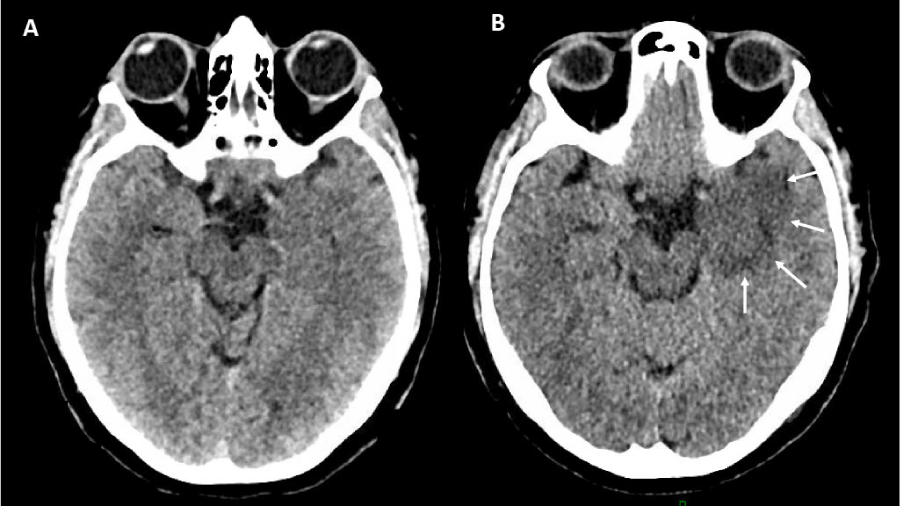 Figure 1: Timing of mesio-temporal edema onset: Comparison between CT on day of epileptic seizure (left) vs. 3 days post-seizure (right).
View Figure 1
Figure 1: Timing of mesio-temporal edema onset: Comparison between CT on day of epileptic seizure (left) vs. 3 days post-seizure (right).
View Figure 1
MRI revealed a large hyperintense area on FLAIR and T2-weighted images in the left mesio-temporal region, extending to the cerebral peduncle, with swelling of the parenchyma. Within this area, numerous vascular structures and hemosiderin deposits were observed, consistent with a DAVF. The expansive parenchymal changes observed were atypical for a chronic reaction to a vascular malformation, raising concerns about an underlying glial tumor (Figure 2).
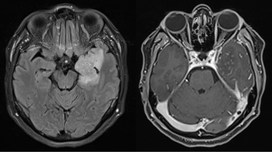 Figure 2: FLAIR and Gadolinium-enhanced t1 sequences from initial MRI.
View Figure 2
Figure 2: FLAIR and Gadolinium-enhanced t1 sequences from initial MRI.
View Figure 2
Arteriography confirmed the presence of a DAVF supplied by various arterial feeders, including the left occipital artery, the left middle meningeal artery, and the ascending pharyngeal artery. The drainage occurs via an excluded venous segment of the left transverse sinus (due to thrombosis of the sigmoid segment) and ultimately through arterialized left cortical veins, which reflux into the superior sagittal sinus (Figure 3, left). The DAVF was classified as type II according to the Borden classification system.
To investigate the possibility of an underlying tumor, a Methionine PET-CT was performed, revealing low to moderate metabolic activity in the affected area, with a maximum SUV of 2.6 in the left thalamus. In the context of post-ictal edema, which typically shows hypoactivity, these findings were consistent with a low-grade glial process.
A complete embolization was performed a few days later using the pressure Cooker technique at the level of the middle meningeal artery. The diseased venous segment was treated with 2.2 ml of Squid 12. Angiographic follow-up confirmed the complete exclusion of the fistula (Figure 3).
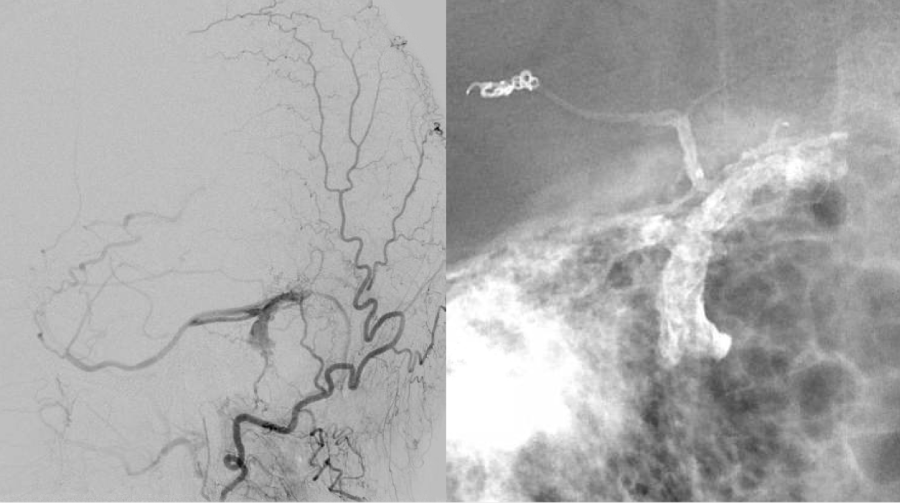 Figure 3: Pre- and post-embolization angiography of a left transverse sinus DAVF: Pre-embolization early venous opacification via arterial feeders from the occipital artery, middle meningeal artery, and extradural vertebral V4 branch (left), and complete obliteration of the pathological venous segment post-embolization (right).
View Figure 3
Figure 3: Pre- and post-embolization angiography of a left transverse sinus DAVF: Pre-embolization early venous opacification via arterial feeders from the occipital artery, middle meningeal artery, and extradural vertebral V4 branch (left), and complete obliteration of the pathological venous segment post-embolization (right).
View Figure 3
Following a multidisciplinary discussion between neuro-oncology and neuro-vascular teams, it was decided to prioritize treatment of the fistula, with close radiological and clinical follow-up. Serial MRIs over the subsequent three months showed progressive and near-complete resolution of the cerebral parenchymal changes (Figure 4), ruling out the initial suspicion of an associated tumor.
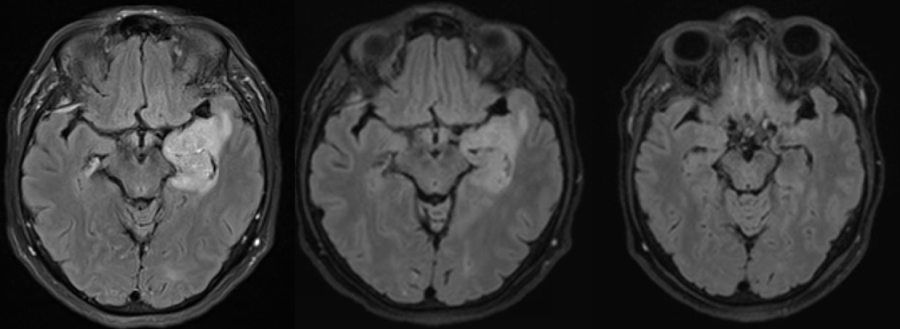 Figure 4: FLAIR sequence evolution: at diagnosis (left), one week post-endovascular treatment (center), and six weeks post-treatment (right).
View Figure 4
Figure 4: FLAIR sequence evolution: at diagnosis (left), one week post-endovascular treatment (center), and six weeks post-treatment (right).
View Figure 4
Intracranial DAVFs are relatively rare entities, representing about 15% of all cerebrovascular malformations. They are generally regarded as acquired conditions, although their precise origin remains uncertain, and correspond to abnormal direct shunts between dural arteries and one or more draining dural veins or sinuses, without an intervening capillary bed [1,2].
Clinical manifestations depend largely on the site and venous drainage pattern, often classified using the Borden or Cognard systems. The presence of cortical veinous reflux (CVR) is key to predicting the future risk of adverse neurological events.
Typical presenting symptoms of a DAVF include pulsatile tinnitus, headache, proptosis, or neurological deficits linked to intracranial hemorrhage [3]. More rarely, patients may develop progressive cognitive decline or Parkinsonian features secondary to chronic hypoperfusion of the cortex or basal ganglia [4].
In this relatively unusual case, the patient experienced temporal lobe seizures related to the uncommon mesio-temporal location of the fistula. These seizures were primarily driven by the irritative potential of the lesion itself, in the absence of hemorrhage or initial edema. Notably, no edema was observed on the first CT scan and appears to have developed subsequently.
Prior research has demonstrated that transient focal MRI changes following status epilepticus (SE) can be attributed to vasogenic or cytotoxic edema, although the exact pathophysiological mechanisms remain debated. Vasogenic edema likely arises from increased permeability due to blood-brain barrier disruption after SE, whereas cytotoxic edema may stem from neuronal injury caused by inflammation, hypoxia, or metabolic dysfunction. Such edema is usually transient and tends to resolve within months [5-7].
In this case, we postulate that the rapidly developing edema was the result of combined effects of chronic venous congestion and SE-related edema. The timing suggests that seizure-induced inflammatory and metabolic disturbances increased blood-brain barrier permeability, thereby worsening a preexisting vulnerability due to venous congestion.
Reports describing severe edema linked to cerebral DAVFs are scarce [8-10], since most of these lesions drain into dural venous sinuses or large cortical veins, which generally accommodate increased flow without causing major venous congestion in adjacent brain tissue. Conversely, such congestive edema is well described in spinal DAVFs, as spinal veins have a more limited capacity to manage elevated flow.
Some recent publications suggest that pial venous reflux (PVR) has a stronger association with hemorrhage or edema than CVR in DAVF patients, a pattern reminiscent of spinal DAVFs [11]. This reflux is usually detectable only in very late venous phases and may not be visible on standard angiography or 3T MRI. Unfortunately, it was not specifically assessed in our case.
11C-methionine PET can help distinguish malignant neoplasms from benign or non-neoplastic lesions by measuring 11C-methionine uptake, which reflects amino-acid transport and protein synthesis linked to cell proliferation. Malignant tumors typically demonstrate elevated uptake (T/N ratio > 1.5), while benign lesions such as DAVFs usually display normal or reduced methionine uptake [5]. In our patient, Methionine-PET revealed moderately increased uptake (SUV max > 2.6) in the region of MRI abnormalities, similar to a low-grade glioma.
In addition, the DAVF in this case was associated with hemosiderin deposition, likely reflecting focal blood-brain barrier compromise and weakened tight junctions, which may influence methionine accumulation. Prior studies have also indicated that reactive gliosis and inflammatory changes can alter methionine uptake [12,7].
This case highlights the complexities of diagnosing and managing dural arterio-venous fistulas, especially when confronted with atypical symptoms and imaging findings that may suggest an underlying pathology. This unusual presentation underscores the importance of thorough evaluation and multidisciplinary collaboration in such cases.
The successful embolization of the DAVF, which led to the resolution of symptoms and cerebral parenchymal changes on follow-up imaging, highlights the significance of timely intervention. This case contributes to a better understanding of the complex relationship between DAVFs, associated parenchymal changes, and their neurological implications.