
Older adults frequently present with atypical or blunted symptoms of infection, complicating diagnosis and management. This report describes two elderly patients with complex urosepsis-one with a prostate abscess and the other with a ureteric stone—who initially appeared clinically stable. Despite downward-trending septic markers, persistent clinical suspicion prompted further investigations that revealed significant underlying pathology. These cases illustrate the importance of holistic assessment, proactive imaging, and patient advocacy in managing infections among frail adults, underscoring that apparent improvement should not preclude further evaluation.
Complex urosepsis, Septic emboli, Prostate abscess, Streptococcus agalactiae, Ureteric stone
Urosepsis in older adults presents diagnostic challenges due to age-related physiological changes and multimorbidity. Classical signs of infection such as fever, dysuria, or flank pain may be absent or muted in this population, delaying targeted management and potentially worsening outcomes. Frailty, altered immunity, and polypharmacy further complicate the clinical picture. Current guidelines emphasize the importance of individualized, comprehensive assessment that extends beyond laboratory parameters and standard clinical cues [1].
This case series demonstrates how persistence in clinical advocacy and timely imaging led to identification of concealed urinary tract pathology, preventing deterioration despite apparent clinical stability.
An 89-year-old gentleman who lives alone, is functionally independent with a Clinical Frailty Scale (CFS) score of 4. His medical history includes diabetes mellitus, hypertension, and dyslipidaemia. His old medications include; Metformin 850 mg once daily and Simvastatin 10 mg once daily; however, he was non-adherent and had been lost to follow-up.
He was admitted after a syncopal episode in the toilet. He reported squatting during a bowel movement and, upon attempting to stand, experienced sudden weakness in both legs, leaving him unable to rise. He remained seated for about two hours before trying to stand again. He also reported a six-month history of altered bowel habits, including intermittent loose stools and vomiting, especially when unable to pass stool.
Investigations revealed iron deficiency anaemia with an initial suspicion of gastrointestinal malignancy. His haemoglobin dropped from 7.2 g/dL to 6.3 g/dL pre-transfusion and improved to 8.0 g/dL post-transfusion. Red cell indices showed an MCV of 76 fL, MCH of 23.8 pg, and MCHC of 314 g/L. Serum iron was low at 1.7 µmol/L, with transferrin saturation at 5%. Abdominal X-ray showed a dilated bowel measuring 7.3 cm (Figure 1). Tumour markers revealed CA19-9 at 54 U/mL and CEA at 2.2 ng/mL. Faecal occult blood testing was positive (Table 1).
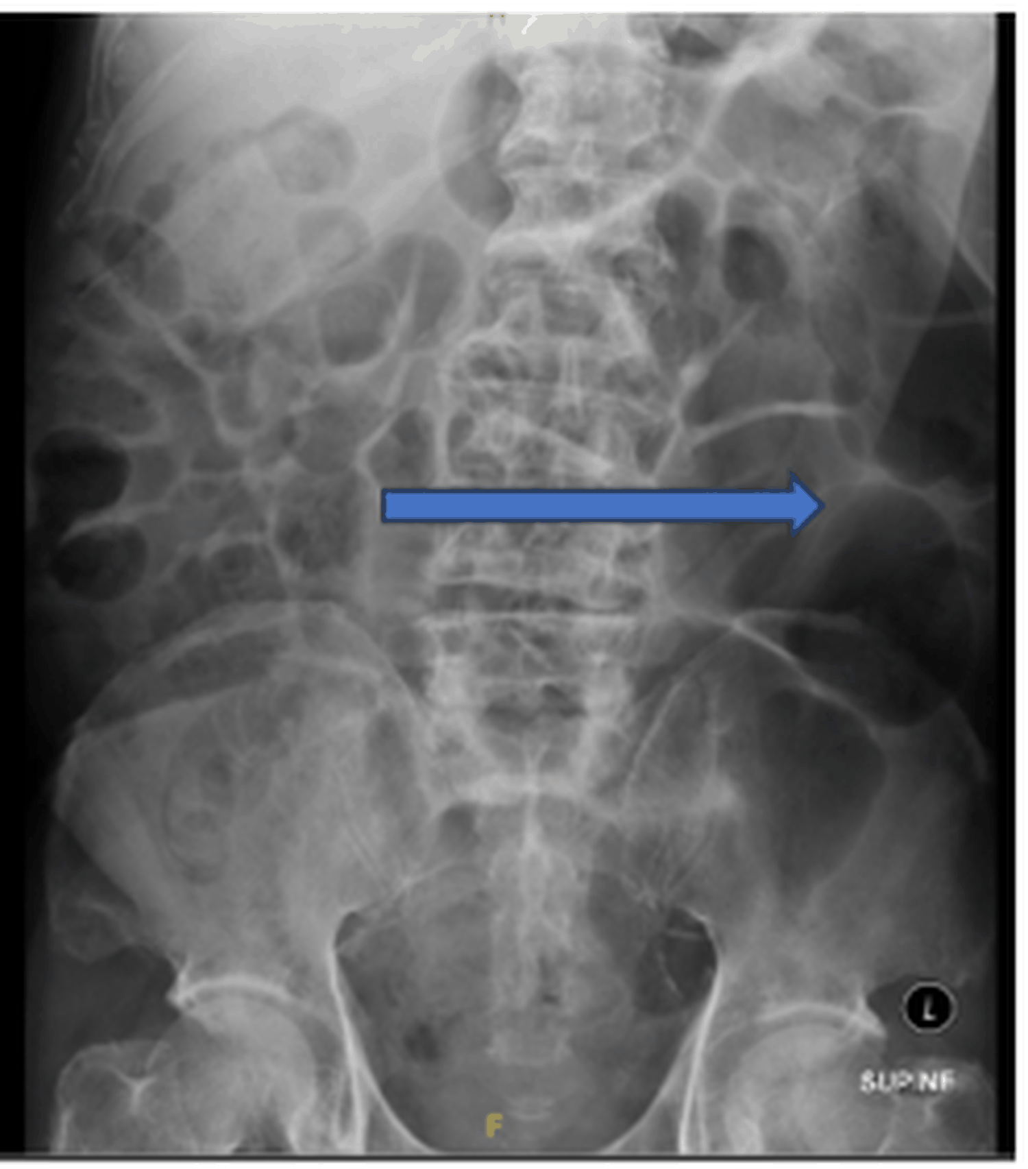 Figure 1: Abdominal radiograph showed dilated bowel.
View Figure 1
Figure 1: Abdominal radiograph showed dilated bowel.
View Figure 1
Table 1: Summary of investigations. View Table 1
A contrast-enhanced CT scan of the abdomen and pelvis on 12/6/25 showed an enlarged prostate measuring 4.5 x 5.6 x 4.3 cm (volume 56.3 mL) with multiple ill-defined hypodense lesions; the largest at the apex measured 1.0 x 1.0 x 0.9 cm (Figure 2). There was no significant lymphadenopathy or intestinal obstruction. Splenomegaly with possible infarcts was noted.
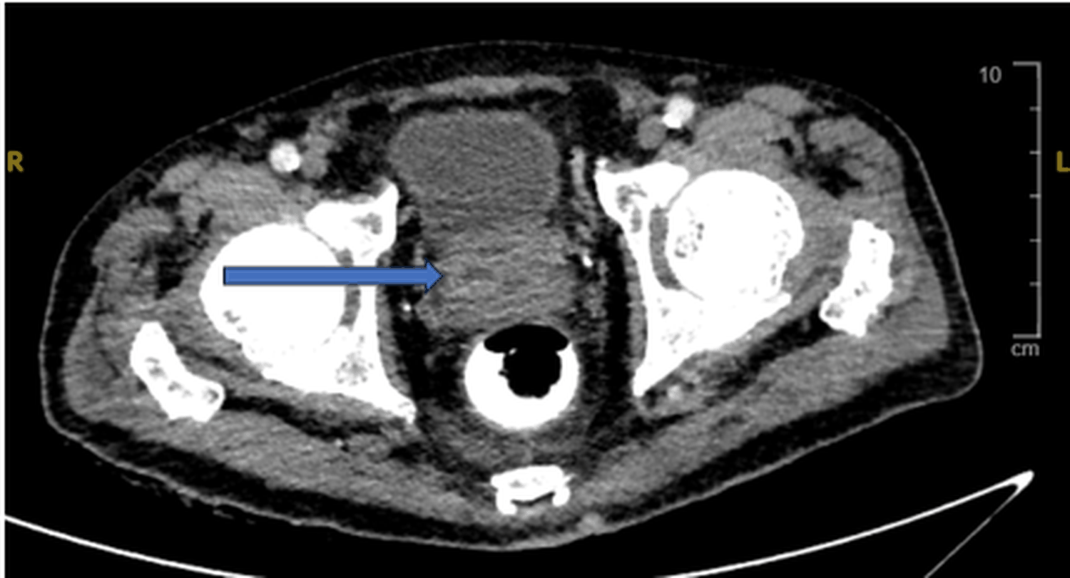 Figure 2: CT scan of the abdomen showed an enlarged prostate.
View Figure 2
Figure 2: CT scan of the abdomen showed an enlarged prostate.
View Figure 2
He underwent esophagogastroduodenoscopy (OGDS) on 13/6/25, revealing D1 duodenitis and a fundal polyp; two biopsies were taken. The rapid urease test was negative. Colonoscopy was limited by poor bowel preparation but reached the transverse colon with normal findings.
During admission, he was diagnosed with E. coli urosepsis complicated by a prostate abscess and possible splenic infarcts, suggesting septic emboli. Urine analysis was positive for nitrites, leukocytes (3+), and blood (3+), although the sample was likely from a catheter. His septic markers trend downward, and he remained clinically well. kidneys, ureters, and bladder (KUB) showed no stones. Blood cultures were negative. The infectious diseases team reviewed him, and intravenous Augmentin was restarted.
A 76-year-old lady with a Clinical Frailty Scale (CFS) score of 4. She lives alone; her son resides in Australia, and her daughter lives in Kota Bharu. Her medical history includes diabetes mellitus, osteoporosis, non-alcoholic steatohepatitis (NASH), asthma, hypertension, and ischemic heart disease.
Her previous medications included Mixtard insulin 24/36 units and Metformin 1 gram twice daily. About one month prior to admission, during a visit to her son in Australia, she did not administer Mixtard regularly and was instead prescribed Metformin and Empagliflozin. Her most recent HbA1c was 7.6%.
She was admitted after a two-day history of fever. When her children could not reach her, a neighbour arranged for a locksmith to enter her home. She was found confused and soiled with urine and faeces on the floor.
On admission, she was clinically dry, weak and delirious with tender lower abdomen. Laboratory results showed blood glucose of 25 mmol/L, lactate rising from 2.5 to 2.85 mmol/L, and ketones at 0.6 mmol/L. Urine analysis showed leukocytes 3+ and negative nitrites. Creatinine was 141 µmol/L, urea 6.6 mmol/L, CRP elevated at 222 mg/L, platelets 202 x10 9 /L, haemoglobin 12.2 g/dL, and INR 1.22. She was treated for urinary tract infection and uncontrolled diabetes complicated by lactic acidosis.
Blood and urine cultures grew Streptococcus agalactiae bacteraemia. She was initially treated with intravenous Rocephin for four days, then switched to benzylpenicillin. While in ward, she had a fever spike but otherwise maintained a good appetite, not delirious, participated in physiotherapy activities, and was able to ambulate to the toilet independently. Her CRP decreased from 155 to 26 mg/L, and her renal profile further improved.
Subsequent abdominal ultrasound revealed a right ureteric stone (Figure 3), which was then stented. During the RPG stent procedure, urine cultures grew Pseudomonas veronii. The infectious diseases team recommended completing one week of Fortum (ceftazidime) therapy.
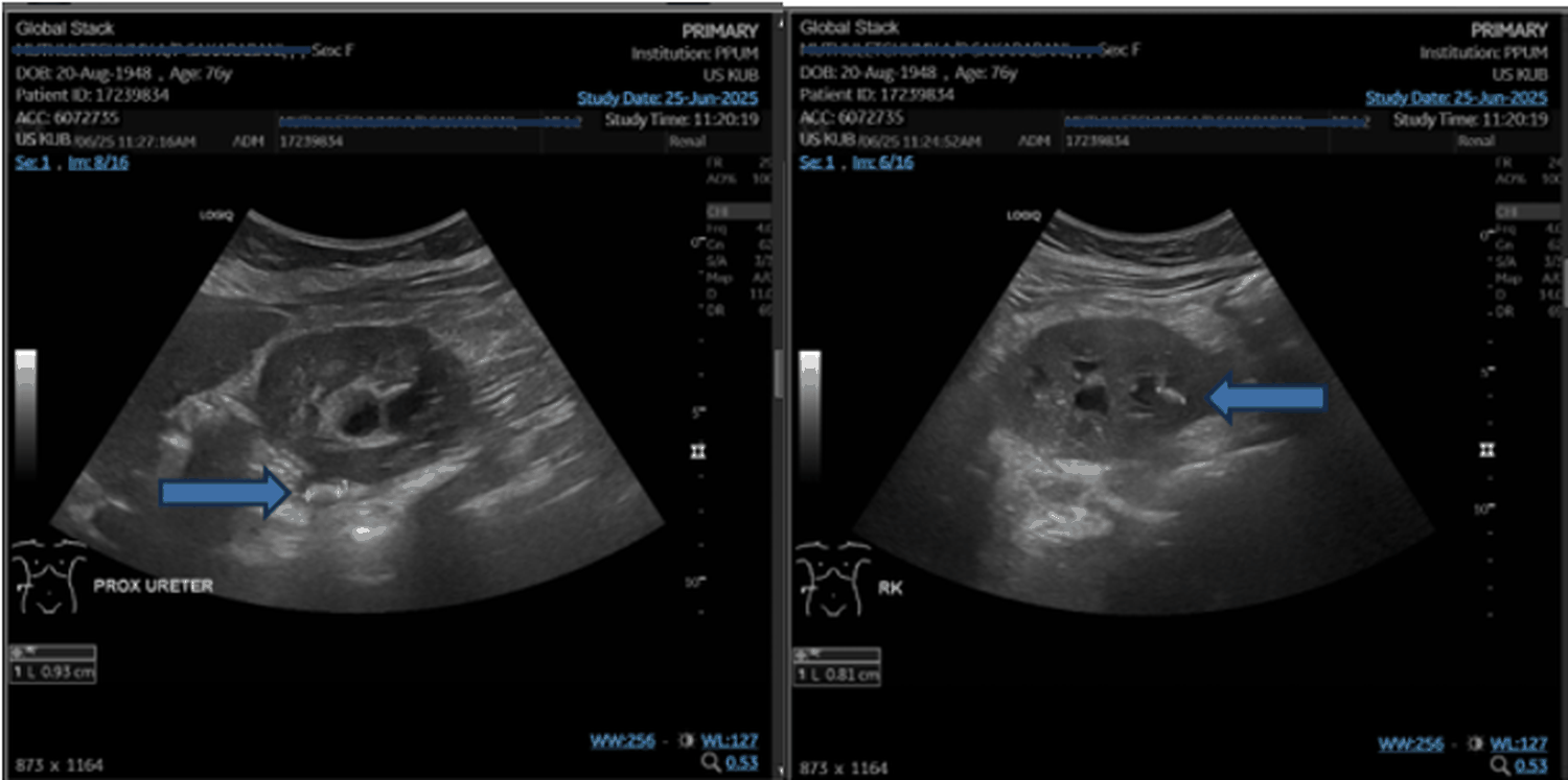 Figure 3: Right proximal ureter calculus causing moderate hydronephrosis and right renal calculus.
View Figure 3
Figure 3: Right proximal ureter calculus causing moderate hydronephrosis and right renal calculus.
View Figure 3
Both cases highlight the complexity of diagnosing and managing urosepsis in older adults. Despite apparent clinical improvement and resolving inflammatory markers, each patient harboured significant underlying pathology. These cases illustrate several shared themes and learning principles relevant to geriatric infection management.
Neither patient presented with classical urinary symptoms. The first case manifested as syncope and non-specific bowel complaints, while the second involved delirium and dehydration-both common atypical infection presentations in the elderly [2]. Such manifestations reflect diminished physiological reserve and altered host response to infection [3].
In both cases, clinical stability and biochemical improvement could have prematurely ended diagnostic evaluation. However, subtle ongoing abnormalities prompted continued assessment, revealing a prostate abscess in Case 1 and a ureteric obstruction in Case 2. This underscores that in older adults, apparent recovery may mask incomplete source control.
Persistent advocacy for imaging was the turning point in both scenarios. Although a conservative approach might have sufficed initially, imaging uncovered reversible anatomic causes for ongoing infection. This aligns with recommendations emphasizing timely imaging for suspected complicated urinary infections, particularly when sepsis resolves atypically or source control is uncertain [4].
Shared principle - patient advocacy through comprehensive reassessment.
The unifying principle across both cases is patient-centred advocacy: clinical improvement should not end diagnostic curiosity. For frail or cognitively impaired patients, advocacy extends beyond routine monitoring to proactive investigation. Such practice embodies the evolving definition of patient advocacy—not limited to consent facilitation or safety, but encompassing the pursuit of diagnostic completeness and therapeutic adequacy [5].
Each case presents distinct microbiological and anatomical complexity. E. coli bacteriuria commonly complicates prostate abscesses, whereas Streptococcus agalactiae and Pseudomonas co-isolation in Case 2 reflects polymicrobial infection risk after instrumentation. Their distinct organisms and anatomical involvement underline that achieving genuine source control in elderly patients requires case-by-case tailoring, not standardized antibiotic duration or empirical regimens.
Ultimately, these cases affirm that infection management in older adults is dynamic and contextual. Core geriatrics principles-individualized care, comprehensive assessment, and interprofessional collaboration—remain central to achieving both recovery and preservation of autonomy.
These cases reinforce the principle that infection management in older adults must extend beyond resolving sepsis. Clinical vigilance, patient advocacy, and individualized reassessment are essential, as normalizing laboratory results may conceal unresolved sources. Atypical presentations and blunted inflammatory responses demand integration of functional, cognitive, and imaging assessments to ensure adequate diagnosis and lasting recovery.