
Background: Asbestiform fibers and cleavage fragments of the same mineral have different dimensional distributions. The paper explores how dimensional differences due to mineral habit affect their concentration in human lungs after exposure.
Methods: Dimensional characteristics of asbestiform amosite and non-asbestiform grunerite were used in this study. The Rasmuson-Roggli method was utilized to predict average lung fiber concentration based on cumulative exposure. The Deposition Selection Ratio (DSR), which is a function of length and width of fibers, was estimated from dimensional data in human lungs, as a correction factor to average exposure. A hypothetical exposure that averaged 4 f/cc-years of asbestiform fibers and of cleavage fragments (standard deviation 1 f/cc-years) longer than 5 µm was assumed.
Results: The method predicted 95th percentiles of 0.47 mf/g of dry lung tissue for asbestiform particles vs. 0.06 mf/g of dry lung tissue for cleavage fragments. For cleavage fragments, the concentrations were shown to be below published concentrations of amphiboles in the general population (not expected to be related to elevated risk of cancer).
Conclusions: Risk analysis demonstrated that asbestiform fibers are about an order of magnitude more likely to be deposited in lungs for prolonged periods, and mesothelioma risk is about two orders of magnitude higher for asbestiform fibers per unit of accumulated lung dose.
Asbestiform fibers, cleavage fragments, mesothelioma, lung concentrations, Deposition Selection Ratio (DSR)
Elongate mineral particles (EMPs) produce adverse effects in humans by penetrating the respiratory system through breathing and creating unbalanced processes of deposition, clearance, and toxicological impact on the levels of cells and tissues (Attanoos, Allen, 2014) [1]. Elongationis a main driver of toxicity for asbestos and asbestiform particles, with clear differentiation between long, thin particles producing such outcomes as mesothelioma and particles with different shapes that are not mesotheliomagenic (Pass, et al. 2005) [2]. Recent publications significantly advance the scientific basis of EMP toxicology which was demonstrated to be closely related to their mineralogical characteristics [Wylie, Korchevskiy (2023); Wylie, Korchevskiy (2025); Korchevskiy, Wylie (2025); Korchevskiy, Wylie (2025); Korchevskiy, Wylie (2025)] [3-7].
Morphology of fibers is one of the parameters that has been underappreciated in toxicological studies for decades. However, the type of geological growth of asbestiform minerals creates a clear differentiation for fibers with characteristics of commercial asbestos vs. so called “cleavage fragments” that are produced by mechanical forces from specific types of minerals but do not possess dimensions, strength, and other parameters that are associated with carcinogenic effects (Gamble, Gibbs, 2008; Wylie et al., 2022) [8,9]. Biologically inert cleavage fragments can be inhalable dust, but they are not carcinogenic asbestos (Addison, McConnell, 2008) [10].
It is well known that concentrations of asbestos fibers in lungs are predictive of mesothelioma mortality (Rasmuson et al., 2014; Gilham et al., 2016) [11,12]. However, there is a lack of information about the process of deposition and clearance of cleavage fragments in human lungs. Korchevskiy, et al. (2025) [13] suggested that dimensional parameters of elongate particles drive their propensity to be deposited in lungs. It is important to test this hypothesis in conjunction with methods for prediction of lung concentrations in relationship to cumulative exposure.
To propose a method for estimating concentrations of elongate particles in human lungs depending on the cumulative exposure to fibers and their dimensional distribution.
To compare the predicted lung concentrations for particles belonging to different habits (asbestiform vs. non-asbestiform, or cleavage fragments).
To determine the implications for carcinogenic risk assessment of the difference between fiber concentrations in lungs for asbestiform fibers and cleavage fragments.
Asbestiform amosite along with its non-asbestiform (fragments) variety referred to by their mineral names grunerite were selected for the analysis as representative examples of commercial asbestos and corresponding cleavage fragments. The discussion about the choice of amosite and non-asbestiform grunerite for dimensional analysis along with original dimensional data for these minerals can be found in Korchevskiy, Wylie (2025) [7].
Estimates of fiber concentrations in lungs based on cumulative exposure for amosite was developed using the Rasmuson-Roggli non-linear regression (Rasmuson et al., 2014) [11].
The relationship between cumulative exposure to amosite fibers with length > 5 µm and concentration of fibers in lungs was determined as:
CE = 10^((Log 10 (LFSEM(wet))-3.65242)/0.83422), (Equation 1)
where CE is the cumulative exposure to amphibole fibers longer than 5 µm (in f/cc-years), and LFSEM(wet) is the lung concentration of amphibole fibers by Scanning Electron Microscopy (SEM) (f/g of wet tissue).
The relationship between fiber concentrations in dry and wet tissue was assumed to be linear, with the coefficient of about 10 (Korchevskiy, Wylie, 2022): [14]
LF SEM(dry) = 10 x LF SEM(wet) (Equation 2)
Also, we used a linear relationship for the derivation of concentration projected by transmission electron microscopy (TEM) that is supposed to be better suitable for estimates of dimensional parameters of fibers (Korchevskiy, Wylie, 2026): [15,16]
LF SEM(wet) = K x LF TEM(wet) (Equation 3)
We derived the coefficient K from the comparison of dimensional distributions of amosite data in the dimensional database (Korchevskiy, Wylie, 2025 Supplemental material) [7] by SEM vs. TEM, which shows that the thinnest fibers are often ignored by SEM but counted by TEM.
If t is a fraction of fibers with width ≤ 0.25 µm by TEM (assumed to be correctly determined), X0 - number of fibers with width ≤ 0.25 µm by SEM and Y0 - number of fibers with width > 0.25 µm by SEM,
we can determine K from the formula
1/K = ((tY0/X0(1-t)+Y0)/(X0+Y0) (Equation 4).
Based on our data on SEM and TEM measurements of amosite samples, we obtained the estimations of t = 0.45, X0 = 668, Y0 = 1790, and from here we estimated K ≈ 0.8.
To account for the difference in deposition and clearance of fibers in human lungs, we used aparameter called deposition selection ratio (DSR), first proposed by Korchevskiy, et al. (2025) [13] which was calculated for each size group following Equation 5:
DSR i = f i (lungs)/f i (exposure), (Equation 5)
Where i - size group,
f i (lungs) - fraction of elongate particles of the group i in lungs, and
f i (exposure) - fraction of elongate particles of the group i in the exposure.
As was shown in Korchevskiy A, (2025) [13], DSR can be approximated by a regression equation from length and width of elongate particles in the following way:
Log 10 (DSR+0.001)=-A+Blog 10 (length)-Clog 10 (width), (Equation 6)
where A, B and C are certain coefficients (that will be evaluated further in this paper based on several considerations).
Korchevskiy, A, (2025) [13] determined that based on human data derived from previously published Pooley and Clark information, the coefficients for the DSR equation can be found as:
A = 1.09
B = 0.53
C = 1.03
(R = 0.62, R 2 = 0.38, p < 0.000001). While the correlation is not very high, Korchevskiy, Attanoos, and Wylie showed that DSR estimates based on the formula are consistent with the observed values for the range of sizes of elongate particles typical for human exposure.
From here, we can determine that:
LFTEM(dry)~ 10^(0.83422Log10(CE)+3.65242)/1000000*10/0.8 x DSR/L = 10^(0.83422Log10(CE)+3.65242)/1000000*10/0.8 x (10^(-A+Blog10(length)-Clog10(width))-0.001)/L (mf/g), (Equation 7)
where L is a central tendency value of DSR for asbestiform elongate particles longer than 5 µm, included in this study. The choice of weight coefficient L as the average between the asbestiform varieties of particles is substantiated by the fact that the Rasmuson-Roggli model was derived for commercial amphiboles. Mathematically, using DSR/L as a coefficient for specific fiber sizes can be confirmed by averaging the lengths and widths of all particles; this yields avalue directly from the equation derived by Rasmuson et al. (2014) [11]. We calculated L as the central tendency value for the amosite dimensional data available in our study.
Using Equation 7, the statistical distribution of lung fiber concentrations can be determined by varying the distributions of CE, length, and width. We performed a statistical simulation with 10,000 iterations to estimate LF TEM(dry) for the exposure scenario described below.
In our study, we modeled CE as a uniform distribution with an average of 4 f/cc-year’s and standard deviation of 1. The level of 4 f/cc-years is conditional, corresponding to the hypothetical 40 years of exposure to 0.1 f/cc, a standard occupational limit for asbestos exposure (OSHA, 1994) [17].
We utilized DSR to determine the size distribution of fibers in human lungs, by applying this size dependent coefficient as a relative probability to each fiber in the original distribution of amosite fibers (as derived from our original data, referenced in Korchevskiy, Wylie, 2025) [7].
To estimate lung concentrations from cumulative exposure, we used exposure distributions for length and width that correspond to available dimensional data for amosite and non-asbestiform grunerite. For this purpose, Monte Carlo simulation was utilized, with CE, length and width changing according to the corresponding distributions, and the results were reported as distributions of lung concentrations according to Equation 7. Only particles with length >5 µm, length/width ≥ 3, a width ≥ 0.05 µm, and width ≤ 3 µm were included in the simulation in order to align with existing models of cumulative exposure derived by Rasmuson-Roggli.
We also estimated the distribution of length and width of elongate particles in lungs, based on the estimated distribution in the exposure, with DSR serving as a weighting factor.
Also, using the calculated predicted lung concentrations, we created a hypothetical visual simulation of asbestiform and non-asbestiform elongate particles as they would appear on a transmission electron microscope (TEM) grid. This visualization compares the probability of detecting elongate particles in human lungs. Because of the relationship between mesothelioma risk and detected concentrations of fibers in lungs, this approach makes it possible to visualize the differences in cancer risks.
The number of particles to be placed on hypothetical TEM grid was found as
Number of particles = LF TEM(dry) /AS,
where AS - typical analytical sensitivity for lung burden analysis by TEM (assumed to be 0.01 mf/g, as in Gilham et al. (2016) [12].
For the dimensional distribution of the particles on the TEM grid, random statistical sampling was performed based on the dimensional distribution of particles in human lungs as determined by transformation according to Equation 6 of this paper.
Then, the particles with the determined distribution of sizes were randomly distributed on the hypothetical TEM grids.
To determine mesothelioma risk per unit of lung dose of elongate particles, we used linear and exponential models from Wylie et al. (2020) [18]. The average of the linear and non-linear estimates can be seen as a robust approximation of the central tendency potency factor. Calculations of lifetime mesothelioma risk for a specific exposure scenario were performed according to the Hodgson, Darnton (2000) [19].
Dimensional characteristics of EMPs for two habits (asbestiform and cleavage fragments) in the exposure (based on dimensional database information) and in the lungs (based on the DSR correction factor) are provided in Table 1.
Asbestiform and non-asbestiform particles in our database differ also by their Pearson index, or the correlation coefficient between log-transformed length and width. For asbestiform particles, Pearson index is 0.133, and for non-asbestiform, 0.484. For predicted distribution of fibers in human lungs, Pearson index is reduced slightly for the asbestiform variety (0.17) but increases for non-asbestiform fraction (0.696).
We estimated that average DSR for amosite with length > 5 µm in our study is 0.89 (standard deviation is 0.71). The central tendency value of 0.89 was used for parameter L in Equation 7.
Notably, DSR for non-asbestiform grunerite is statistically significantly lower than for the asbestiform variety (mean of 0.19, standard deviation 0.11).
The distribution of lung concentrations of elongate particles (mf/g dry weight of lungs) based on Equation 4 from the Rasmuson-Roggli model is provided in Figure 1 (a - asbestiform EMPs, b - non-asbestiform EMPs, c - combined). Only fibers with length > 5 µm, length < 100 µm, length/width ≥ 3, width ≥ 0.05 µm, width ≤ 3 µm are included.
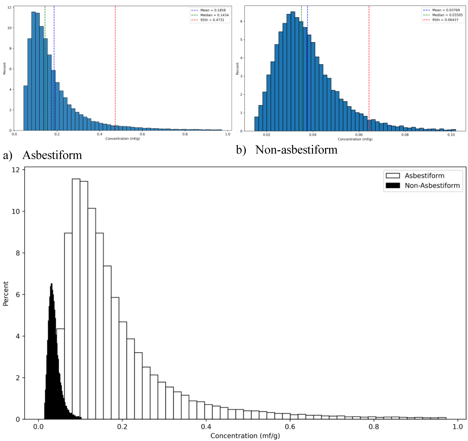 Figure 1: Distribution of lung concentrations of elongate particles (mf/g dry weight of lungs, length > 5 µm, length/width ≥ 3, width ≥ 0.05 µm, width ≤ 3 µm) following cumulative exposure of 4 f/cc-years (standard deviation 1). (a): Asbestiform; (b): Non-asbestiform; (c): Combined histogram (Rasmuson-Roggli model).
View Figure 1
Figure 1: Distribution of lung concentrations of elongate particles (mf/g dry weight of lungs, length > 5 µm, length/width ≥ 3, width ≥ 0.05 µm, width ≤ 3 µm) following cumulative exposure of 4 f/cc-years (standard deviation 1). (a): Asbestiform; (b): Non-asbestiform; (c): Combined histogram (Rasmuson-Roggli model).
View Figure 1
Average concentration of fibers in lungs was found at the level of 0.186 mf/g for asbestiform particles (95 th percentile 0.47) vs. 0.04 mf/g for cleavage fragments (95 th percentile 0.064).
Figure 2 contains a reconstruction of possible TEM microphotographs for lung tissue with asbestiform vs. non-asbestiform population of fibers, conditionally assuming 0.013 mf/g as an analytical sensitivity for the analysis. The grids conditionally contain 100 openings. The leftgrid shows fiber distribution from exposure to asbestiform amosite, and the right grid demonstrates fiber distribution from exposure to non-asbestiform particles (riebeckite). The level of exposure was the same, but, as we can see, the difference in the probability of elongate particles to appear on the grid for a random sample is quite significant.
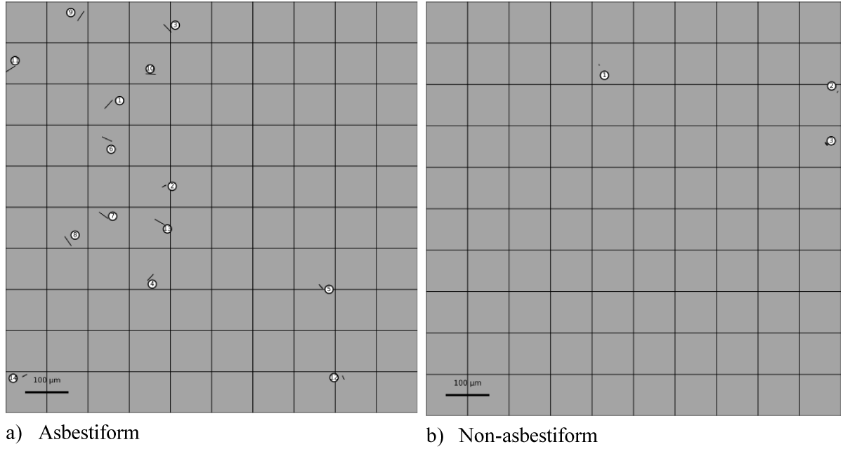 Figure 2: A reconstruction of possible TEM microphotograph for lung tissue with asbestiform vs.non-asbestiform population of fibers, conditionally assuming 0.013 mf/g as an analytical sensitivity for the analysis (cumulative exposure 4 f/cc-years, standard deviation 1). (Themicrophotographs are cleaned from noise of non-elongate particles). Figures a and b demonstrate the TEM grids with random selection of particles according to the size distribution (as in Table 1), with the number of particles corresponding to the concentration of particles in lungs: for asbestiform, 14 particles, for non-asbestiform, 3 particles. The size scale is shown at figures 2a, 2b.
View Figure 2
Figure 2: A reconstruction of possible TEM microphotograph for lung tissue with asbestiform vs.non-asbestiform population of fibers, conditionally assuming 0.013 mf/g as an analytical sensitivity for the analysis (cumulative exposure 4 f/cc-years, standard deviation 1). (Themicrophotographs are cleaned from noise of non-elongate particles). Figures a and b demonstrate the TEM grids with random selection of particles according to the size distribution (as in Table 1), with the number of particles corresponding to the concentration of particles in lungs: for asbestiform, 14 particles, for non-asbestiform, 3 particles. The size scale is shown at figures 2a, 2b.
View Figure 2
Table 1: Length and width of asbestiform and non-asbestiform EMPs (length > 0.5 µm,length < 100 µm, length/width ≥ 3, width ≥ 0.05 µm, width ≤ 3 µm). View Table 1
The population of asbestiform amosite in our study contains 9% EMPA fibers and associated non-asbestiform populations of grunerite contain 0.3% of EMPA fibers. Based on the linear model from Wylie et al. (2020), [18] asbestiform particles in the study would have a mesothelioma potency R M of 0.087%, and non-asbestiform particles would have a negligible R M of < 0 (assuming 0 potency). According to the non-linear model, RM of asbestiform particles will be0.088%, and non-asbestiform 0.00035%. Average values between the two methods would yield 0.087% for asbestiform particles, 0.00017% for the non-asbestiform variety.
Based on risk quantification methods described in Korchevskiy, Rasmuson (2024) [20], exposure to 4 f/cc-years of amosite with a duration of 20 years on average, assuming age of firstexposure as 25 years, would in this case correspond to 211 cases of mesothelioma per 10,000 per lifetime, while the same exposure to grunerite would yield 0.4 cases per 10,000 per lifetime.
According to the Rasmuson-Roggli method (0.186 mf/g for asbestiform, 0.04 mf/g for non-asbestiform particles), we would get 1134 cases of mesothelioma per 10,000 population per 1 mf/g of the dose for asbestiform particles, vs. 10 cases per 10,000 per 1 mf/g of the non-asbestiform variety. It would also comprise about three orders of magnitude of the overall risk difference for the exposure.
Dimensional distribution of elongate mineral particles (EMPs) changes from that of the exposure after particles are inhaled and deposited in lungs. Modeling of particle deposition in lungs shows dependence of the retention fraction on the size of particles (Heyder, 2004; Kim, Hu, 1998) [21,22].The shape of particles affects their deposition according to theoretical models (Asgharian et al., 2018) [23].
Several different studies have attempted to determine the dimensional characteristics of elongate particles accumulated in human lungs. For example, Roggli et al. (1987) [24] demonstrated that there was a progressive increase in mean crocidolite fiber length with time post exposure (P < 0.05), but no significant changes in the diameter of the population of fibers retained in the lung. The authors hypothesized that the tendency for longer fibers to be retained within lung tissue is a characteristic shared by serpentine and amphibole asbestos fibers, and longitudinal splitting with progressive decrease in mean fiber diameter in vivo occurs readily with serpentine fibers. It was unclear if this study measured the relationship between fibers in the exposure vs. long-term retained population or reflected just a change in dimensional characteristics between fibers already deposited. It should be noted that Coffin et al. (1982) [25] demonstrated the same tendency of fiber retention and splitting for amosite and actinolite as in Roggli, et al. (1987) [24]. Coin, et al. (1992) [26] reportedpulmonary deposition, clearance, and translocation of chrysotile asbestos in rats. Adult male ratswere exposed for 3 hours to an aerosol of chrysotile asbestos. Subgroups were sacrificed up to 29 days post exposure and the lungs of the animals fixed. Peripheral and central regions of the left lung were resected, digested, and analyzed for fiber content by scanning electronmicroscopy. Pulmonary deposition did not differ between peripheral and central regions. There was no evidence of translocation of fibers from central to peripheral regions. The average diameter of retained fibers decreased over time. The average length of retained fibers increased over time, consistent with slower clearance of longer fibers and bundle splitting. In particular, fibers with a length ≥ 16 μm were cleared slowly, if at all.
However, the relationship between concentrations of elongate particles with various sizes in thelungs in relationship to their size distribution in the exposure has never been fully established in the literature. Korchevskiy, et al. (2025) [13] noted that published ratios between fiber frequencies in lungs and environmental samples do not correlate with the ones predicted by the US EPA Multiple-Path Particle Dosimetry (MPPD) model (Asgharian , et al., 2018) [23].
The main reason for the differences between MPPD lung concentration prediction and observed relationships between particles in lungs and in the environment may be in the wrong theoretical estimates of the clearance efficiency of particles of various sizes. Long, thin particles appear to be difficult to remove from the lungs because of their complex interaction with macrophages, higher specific surface area (providing better sticking to the biological surfaces), and ability to penetrate deeper into the lung areas where clearance can be hindered (Baranov et al., 2021; Loomis, et al., 2010; Korchevskiy, Wylie, 2026) [27,28,16].
In our study, we used a parameter (Deposition Selection Ratio, or DSR) as a predictor of the“terminal lung deposition” of fibers, understanding this deposition as a steady state process observable after the chronic exposure and lifelong clearance of fibers from the lungs. DSR was evaluated in our recent publication (Korchevskiy, et al. 2025) [13] where we demonstrated that average DSR increases with length and decreases with width of elongate particles. In this study, we demonstrated that there is a correspondence between estimations of DSR based on human data and in a published study on fibers retained by monkeys.
We developed a methodology to predict concentrations of fibers in human lungs that depends on the exposure scenario and length and width of fibers. Two models were used for determination of central tendency lung concentrations. The Rasmuson-Roggli method was derived from observations of SEM lung concentrations in workers vs. retrospectively evaluated cumulative exposure.
Our study confirms that non-asbestiform particles are not expected to be carcinogenic in humans.The difference in carcinogenic potency between asbestiform and non-asbestiform particles is an important fact supported by all available toxicological evidence (Gamble, Gibbs, 2008; Mossman, 2018; Wylie, Korchevskiy, 2023) [8,29,3].
Non-asbestiform elongate particles are predicted to be found in lungs with significantly lower probability than the asbestiform variety. Also, carcinogenicity of cleavage fragments per unit of dose is several orders of magnitude lower than for asbestos fibers. The combination of these two parameters makes cleavage fragments negligible in their carcinogenic potency as has been observed in various studies.
We also utilized visualization to demonstrate a probability of detection of asbestiform fibers vs.cleavage fragments in lung tissue, assuming specific exposure scenario (4 f/cc-years of exposure during 15 to 25 years, after 5 to 15 years from the last exposure). It shows that deposited cleavage fragments would be so rare in human lungs that they will be virtually non-detectable at the grids of a transmission electron microscope (TEM). The associated deposition of asbestiform fibers, to the contrary, will be quite significant and readily found by TEM, with substantial associated risk of mesothelioma.
It is noteworthy that the 95th percentile of lung concentrations for non-asbestiform amphibole exposure in our case does not exceed 100,000 f/g of dry weight, which Visonà, et al. (2025) [30] recently determined as the conditional upper bound of concentrations for amphibole fibers in the lungs of the general population.
It is also expected that shorter, thicker cleavage fragments are less biologically active than asbestiform fibers per unit of the lung dose (Khaliullin, et al. 2020; AG, Korchevskiy, Wylie, 2026) [16, 31,32], so low carcinogenicity of cleavage fragments is a combination of low dose per cumulative exposure and lower toxicity of individual particles accumulated in lungs.
The risk of mesothelioma per a unit of dose was also calculated, by using the risk factor RM as in Hodgson and Darnton (2000) [19] and the relationship between fiber dimensions and mesothelioma potency of amphiboles as determined by Wylie et al. (2020) [18]. Risk analysis demonstrates that asbestiform fibers are about an order of magnitude more likely to be deposited in lungs for prolonged periods, and mesothelioma risk are about two orders of magnitude higher for asbestiform fibers per unit of accumulated lung dose.
Our study has certain uncertainties and limitations. Estimation of DSR potentially needs further in vivo study for confirmation. Nevertheless, the study provides a possible explanation of the differences in distributions of elongate particles in lungs that depend on dimensional parameters. Elongate mineral particles accumulated in lungs show a clear pattern of the asbestiform variety; as can be seen, it is not just a result of prevailing exposure to asbestiform vs. non-asbestiform particles for specific trades, but rather an active selection process for long, thin particles, which are expected to remain in lungs of exposed people for prolonged periods.
1. A modeling approach can be proposed for reconstruction of fiber lung concentration based on cumulative exposure and dimensional distribution of fibers.
2. Asbestiform fibers and non-asbestiform particles (or cleavage fragments) have significantly different dimensional distribution that would drive different behavior in human lungs.
3. With identical cumulative exposure, it was demonstrated that predicted lung concentrations of asbestiform fibers and cleavage fragments would differ at several orders of magnitude.
4. The reconstruction of possible TEM microscopy grids representative of projected fiber concentrations shows that non-asbestiform fibers would be counted at significantly lower proportion, reflecting the difference of the associated carcinogenic risk levels.
Ann Wylie and Andrey Korchevskiy serve as members of the NSSGA Scientific AdvisoryBoard. Andrey Korchevskiy is a coordinator of several scientific projects organized and financed by NSSGA. Ann Wylie was never compensated for her participation in NSSGA-supported scientific projects. The content of this paper was never discussed with NSSGA. No funding was provided by NSSGA for writing the paper. Ann Wylie has served as an expert witness and consultant in litigation involving asbestos.
This research did not receive any specific grant from funding agencies in the public, commercial, or not-for-profit sectors.
The data is available from the authors by reasonable request.
Andrey Korchevskiy (corresponding author): Conceptualization; Investigation; Datacuration; Methodology; Project administration; Software; Supervision; Writing. Arseniy Korchevskiy: Conceptualization; Data curation; Formal analysis; Investigation;methodology; Project administration; Supervision; Validation; Visualization: Writing. AnnWylie: Conceptualization; Data curation; Formal analysis; Investigation; Methodology; Project administration; Supervision; Validation; Visualization: Writing.
The authors acknowledge Debbie Vaughan (Chemistry & Industrial Hygiene, Inc.) fortechnical support in organizing and processing the database information and preparing the paper for publication.