
Tropical pyomyositis is a deep suppurative infection of skeletal muscle, classically described in tropical regions but increasingly reported in patients with metabolic or immune comorbidities. Panton Valentine leukocidin (PVL) producing Staphylococcus aureus strains are strongly associated with severe community acquired skin, soft tissue, and musculoskeletal infections, particularly in African settings where PVL prevalence is markedly higher than in Europe. A 48 year old woman with poorly controlled type 2 diabetes presented with a five week history of progressive, painful swelling of the left leg and fever, without trauma or cutaneous portal of entry. Contrast enhanced CT revealed extensive, multiloculated intra and intermuscular collections involving the entire posterior compartment of the leg. Urgent combined medial and lateral surgical approaches were performed, with wide fasciotomies, thorough debridement of necrotic muscle, compartment decompression, and intraoperative sampling, followed by negative pressure wound therapy. Microbiological work up identified a methicillin susceptible, PVL positive S. aureus strain, with full susceptibility to clindamycin and linezolid; PVL genes (lukS PV/lukF PV) were detected by polymerase chain reaction on the bacterial isolate. Intravenous vancomycin plus clindamycin was started empirically, then de escalated to targeted oral linezolid for two additional weeks, for a total antibiotic duration of four weeks, in line with current recommendations for PVL associated deep infections. The patient had a favorable course, with progressive normalization of inflammatory markers, no need for reoperation, delayed skin grafting, and full functional recovery of the limb. This case illustrates the importance of early imaging, systematic microbiological characterization including PVL testing, aggressive compartment oriented debridement with adjunctive negative pressure wound therapy, and PVL suppressive antibiotic regimens in diabetic patients with extensive tropical pyomyositis.
Pyomyositis, Staphylococcus aureus, Panton‑Valentine leucocidin, Diabetes mellitus, Negative pressure wound therapy
Tropical pyomyositis is a primary bacterial infection of skeletal muscle leading to single or multiple abscesses, traditionally endemic in tropical regions but increasingly reported in temperate climates and in patients with comorbidities such as diabetes, HIV infection, or intravenous drug use. The dominant pathogen is Staphylococcus aureus , and a substantial proportion of cases in Africa are caused by Panton‑Valentine leukocidin (PVL)-producing strains, which are associated with necrotizing soft‑tissue infections and severe invasive disease [1,2]. PVL‑positive S. aureus lineages, particularly clonal complex CC121, account for the vast majority of pyomyositis cases in some pediatric tropical cohorts, strongly suggesting a specific muscle tropism of PVL‑encoding phages. Diabetes mellitus is increasingly recognized as a major host factor in pyomyositis, with recent series reporting diabetic comorbidity in up to one‑third of patients, likely due to impaired neutrophil function, microangiopathy, and altered local defenses. This report describes a severe, multiloculated PVL‑positive S. aureus pyomyositis of the leg in a poorly controlled diabetic adult, with emphasis on the microbiological work‑up, rationale for PVL‑targeted antibiotic therapy, and detailed surgical strategy including wide fasciotomies and negative pressure wound therapy.
A 48‑year‑old woman with a 10‑year history of poorly controlled type 2 diabetes mellitus, treated with metformin and without known chronic complications, presented with a five‑week history of progressive swelling and pain of the left leg associated with documented fever up to 38.7°C and diffuse myalgia. There was no recent history of trauma, strenuous exercise, intramuscular injections, skin lesions, or foreign travel, and no prior episodes of soft‑tissue infection or abscess. Three weeks before admission, she had received oral amoxicillin-clavulanate for presumed cellulitis prescribed by her general practitioner, without clinical improvement. On admission, she was febrile (38.5°C) and hemodynamically stable, with a diffusely swollen left leg, predominantly in the posterior calf, tense and severely tender on deep palpation, but without erythema, warmth, or skin breakdown. Knee flexion was limited by pain, but there was no knee joint effusion, no fluctuance on superficial examination, and no signs of neurovascular compromise or compartment syndrome. Laboratory tests showed leukocytosis of 14,200/mm³ with 90% neutrophils and elevated C‑reactive protein at 145 mg/L, while creatine phosphokinase and renal function were within normal ranges. Blood cultures taken prior to antibiotics, HIV and hepatitis serologies, and urinalysis were negative, and transthoracic echocardiography showed no endocarditis or valvular abnormalities. Given the subacute course, deep location of pain, and absence of overt cutaneous signs, a contrast‑enhanced CT scan of the left leg was obtained as a first‑line modality, which demonstrated multiple intra‑ and intermuscular, low‑attenuation collections with rim enhancement within the posterior compartment (gastrocnemius, soleus, and deep flexors).
The largest confluent collection measured approximately 9 × 6.5 × 27 cm, extending from the popliteal fossa to the distal calf, without intralesional gas or bone involvement (Figure 1). There was no joint effusion and no evidence of osteomyelitis. In the context of a diabetic patient living in a tropical setting, with compatible clinical features and imaging, a diagnosis of suppurative tropical pyomyositis at the abscess stage was made. Intraoperative specimens were obtained systematically from several sites: purulent material from different loculations and biopsies of macroscopically necrotic muscle and fascia. Direct Gram stain showed numerous Gram‑positive cocci in clusters and abundant neutrophils. Aerobic cultures yielded a heavy growth of S. aureus , while anaerobic cultures remained sterile; no other bacteria were isolated. The isolate was identified as methicillin‑susceptible S. aureus (MSSA) by standard automated methods, and full susceptibility testing was performed, showing susceptibility to oxacillin, clindamycin, rifampicin, linezolid, and trimethoprim–sulfamethoxazole, with resistance to macrolides. Because of the severity, tropical context, and association with deep muscle infection, the isolate was tested for PVL production. Detection of the lukS‑PV and lukF‑PV genes was performed by polymerase chain reaction (PCR) on the bacterial DNA, confirming a PVL‑positive strain.
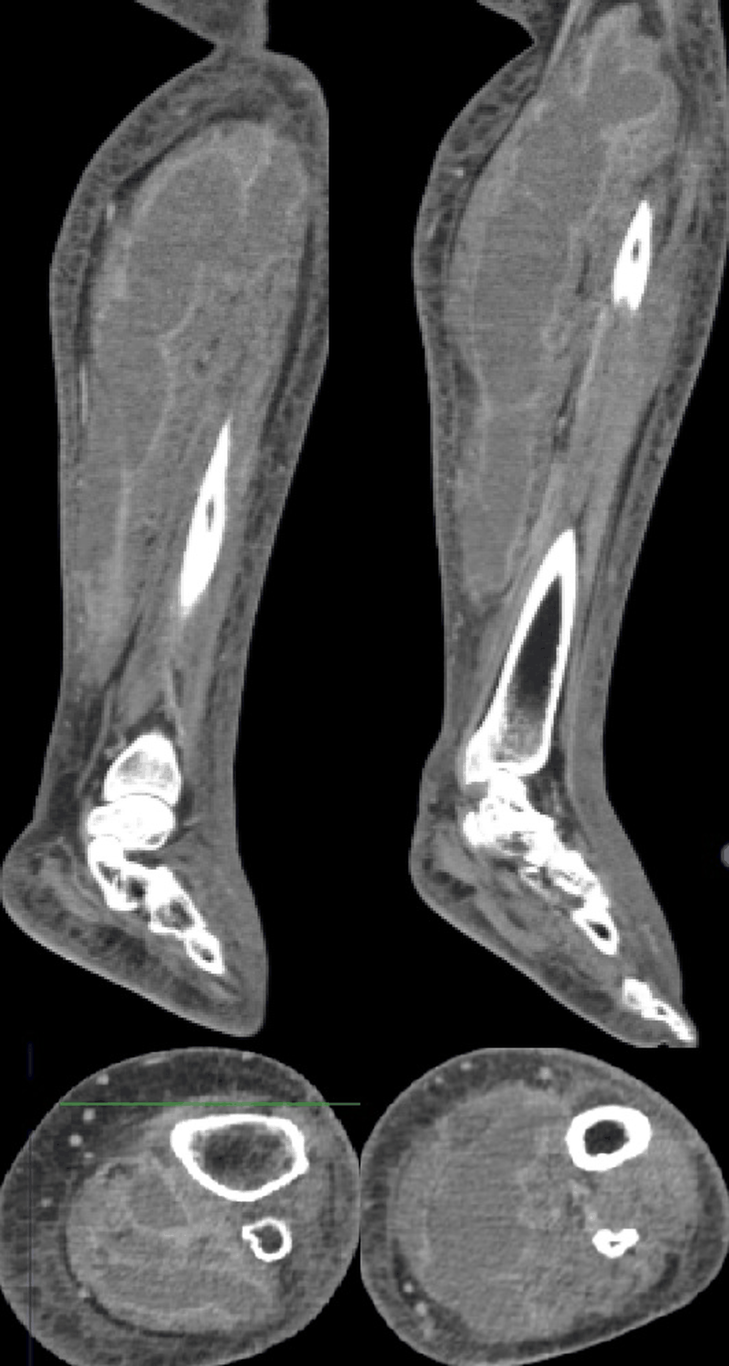 Figure 1: Coronal and axial CT of the left leg showing multiloculated intra-and intermuscular collections in the posterior compartment, forming a 9 × 6.5 × 27 cm abscess without air-fluid levels.
View Figure 1
Figure 1: Coronal and axial CT of the left leg showing multiloculated intra-and intermuscular collections in the posterior compartment, forming a 9 × 6.5 × 27 cm abscess without air-fluid levels.
View Figure 1
No additional virulence or resistance determinants (such as MRSA‑associated SCC mec elements) were identified. Blood cultures remained negative throughout, in keeping with localized deep tissue infection without documented bacteremia. Given the extensive, multiloculated nature of the collections and the risk of progression to septic shock and compartment syndrome, urgent surgical exploration was undertaken within 12 hours of admission. Under general anesthesia and tourniquet control, combined medial and lateral longitudinal incisions were performed to expose the entire posterior compartment and both superficial and deep muscular planes of the leg. Wide fasciotomies of the posterior and lateral compartments were carried out to decompress the muscle groups and allow complete access to the abscess cavities. Intraoperatively, multiple thick‑walled, loculated abscesses separated by fibrous septa were encountered, with areas of grayish, necrotic muscle and diffuse fascial involvement, which is consistent with toxin‑mediated PVL‑associated damage. All loculations were opened and drained, devitalized muscle and fascia were thoroughly debrided, and copious irrigation with isotonic saline was performed until viable, bleeding muscle was observed. The wounds were left open, and negative pressure wound therapy was applied using polyurethane foam dressings connected to a vacuum system set at continuous negative pressure (Figure 2). Postoperatively, the patient was managed in a monitored surgical ward. Serial clinical examinations, daily assessment of pain, limb perfusion, and compartment pressures by palpation, and twice‑weekly laboratory tests (including C‑reactive protein and leukocyte counts) were performed. No residual collections were detected on a follow‑up ultrasound performed at day 7. Wound vac changes were scheduled every 72 hours, combined with gentle passive knee mobilization under sedation to prevent stiffness.
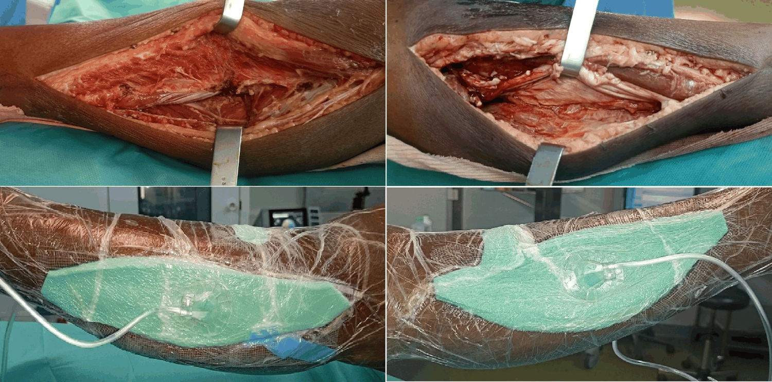 Figure 2: Clinical appearance of lateral and medial leg after wide fasciotomies, debridement, and initiation of negative-pressure wound therapy.
View Figure 2
Figure 2: Clinical appearance of lateral and medial leg after wide fasciotomies, debridement, and initiation of negative-pressure wound therapy.
View Figure 2
The patient required no reoperation, and there were no complications such as deep vein thrombosis or secondary compartment syndrome. Empiric intravenous antibiotic therapy with vancomycin (loading dose then adjusted to achieve therapeutic trough levels) and clindamycin (1.2 g four times daily) was initiated immediately after intraoperative sampling, to cover both MRSA and MSSA and to inhibit PVL and other toxin production. Once cultures confirmed MSSA susceptible to clindamycin and linezolid and PVL positivity, vancomycin was discontinued, and a regimen combining continued IV clindamycin for one week followed by targeted oral linezolid 600 mg twice daily for two additional weeks was adopted, for a total treatment duration of four weeks.
This strategy was chosen to maintain good soft‑tissue penetration and preserve PVL‑suppressive activity while simplifying outpatient management. Inflammatory markers declined steadily, with C‑reactive protein normalizing by week 3, and the patient remained afebrile after day 3. After three weeks of negative pressure wound therapy and satisfactory granulation, delayed split‑thickness skin grafting was performed with uneventful healing (Figure 3). At three‑month follow‑up, the patient was pain‑free, had regained full range of motion of the knee and ankle, and was able to walk without assistance, with no clinical or radiological signs of recurrence.
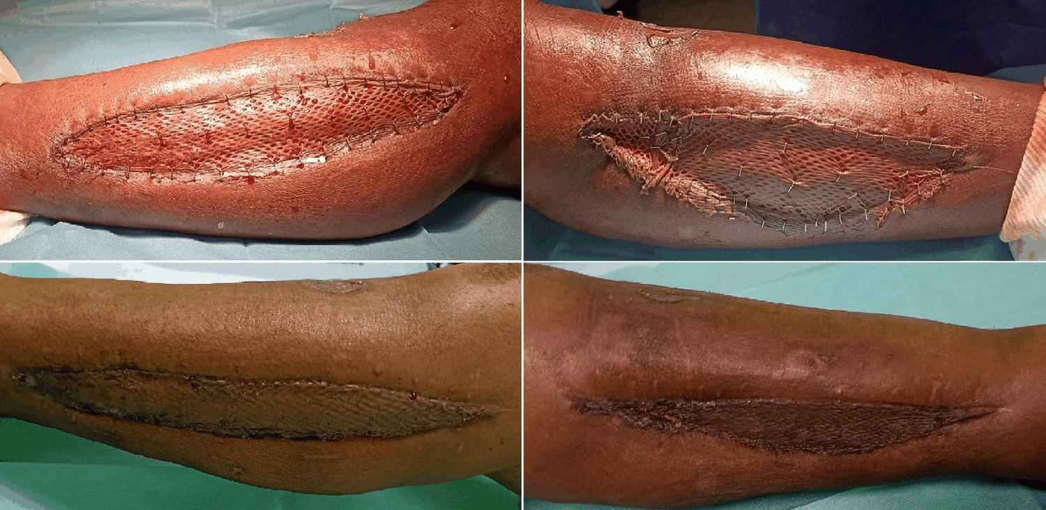 Figure 3: Post-debridement granulation and split-thickness skin graft with satisfactory take at staple removal.
View Figure 3
Figure 3: Post-debridement granulation and split-thickness skin graft with satisfactory take at staple removal.
View Figure 3
Skeletal muscle is normally resistant to bacterial invasion, and several mechanisms have been proposed to explain pyomyositis, including minor trauma, muscle ischemia, nutritional deficiencies, and transient bacteremia [1,2]. In tropical regions, thiamine deficiency and repeated microtrauma have been suggested as co factors that render muscle susceptible to hematogenous seeding by S. aureus [2,3]. PVL is a bicomponent leukotoxin encoded by lukS-PV and lukF-PV that forms pores in neutrophil membranes, leading to cell lysis, massive release of inflammatory mediators, and tissue necrosis [4,5]. In a large Cambodian pediatric cohort, 85% of pyomyositis cases were caused by PVL positive S. aureus of clonal complex CC121, and PVL carriage explained almost all of the bacterial genetic association with pyomyositis compared with asymptomatic carriers, strongly suggesting that PVL is a key determinant of muscle infection in this context [6]. PVL positive S. aureus strains are much more frequent in Africa than in Europe, with reported rates of 45-74% among S. aureus isolates in African settings versus around 3% in European cohorts, which may contribute to the high burden of severe skin, soft tissue, and musculoskeletal infections in tropical regions [7,8]. Diabetes mellitus has emerged as a prominent comorbidity in pyomyositis, with more recent series reporting diabetic patients in approximately 30% of cases, compared with less than 10% in older reports [1,9]. Poor glycemic control impairs neutrophil chemotaxis, phagocytosis, and intracellular killing, alters complement function, and causes microvascular disease and neuropathy, all of which facilitate deep tissue infection and delay recognition of symptoms [10]. In the present case, poorly controlled diabetes may have favored both the initial muscle seeding and the progression of infection despite early empirical oral antibiotics, and likely contributed to the subacute, paucisymptomatic cutaneous presentation [9,10]. Clinically, pyomyositis evolves through three stages: An initial invasive stage with nonspecific pain and low grade fever, a suppurative stage with abscess formation, and a late septic stage with systemic dissemination and metastatic abscesses [1,2].
Diagnostic delay is common, especially when cutaneous signs are absent, as in this patient, and musculoskeletal pain may be misattributed to osteoarticular or neurologic causes [2,11]. CT and MRI are crucial to delineate the extent of intramuscular collections, guide surgical planning, and exclude alternative diagnoses such as necrotizing fasciitis or osteomyelitis [11,12]. In resource limited or emergency settings, CT is often more readily available and can reliably detect multiloculated abscesses and compartment involvement, as in this case [12]. Once suppurative pyomyositis is established, surgical drainage is mandatory and should be extensive in PVL associated disease, where multiloculated abscesses and diffuse fascial involvement are common [5,6]. Involvement of an entire muscle compartment, as in this case, justifies wide exposure through multiple incisions, systematic fasciotomies for decompression and access to deep loculations, meticulous debridement of all devitalized tissues, and generous irrigation [11,13].
Negative pressure wound therapy is particularly valuable in this setting, as it maintains continuous drainage, reduces edema and bacterial load, improves local perfusion, and promotes granulation tissue formation, thereby facilitating delayed closure or grafting [14]. In toxin mediated infections such as PVL positive S. aureus disease, repeated VAC changes also offer opportunities for close monitoring and early detection of residual infection, potentially reducing the need for reoperation [14]. Although evidence is mostly limited to case series and expert opinion, the combination of early, wide debridement and negative pressure wound therapy appears to improve both infectious control and functional outcomes in extensive pyomyositis [13,14]. Antibiotic therapy for PVL associated infections must address both bacterial eradication and toxin suppression [4,5].
International guidelines on PVL positive S. aureus infections recommend initial combination therapy with agents such as clindamycin, linezolid, and rifampicin, which inhibit protein synthesis and reduce PVL and alpha toxin production, rather than relying solely on beta lactams or glycopeptides [5,15]. In severe cases or when MRSA is possible, vancomycin or linezolid may be combined with clindamycin and rifampicin, with subsequent de escalation once susceptibility is known [15]. In the present case, empiric vancomycin plus clindamycin was appropriate pending culture results, and switching to an oral PVL suppressive agent (linezolid) with excellent bioavailability provided effective continuation therapy while facilitating early discharge [5,15]. The four week total duration of antibiotics is consistent with reported regimens for deep muscle abscesses, particularly in diabetic or PVL positive infections, although no randomized data specifically address optimal duration in pyomyositis [1,11]. Antimicrobial stewardship remains important, and the decision to use linezolid should balance the need for toxin suppression and oral step down against potential hematologic toxicity, which mandates close monitoring [15]. This case provides a detailed description of PVL positive MSSA tropical pyomyositis of the leg in an uncontrolled diabetic adult, integrating microbiological characterization, rational use of PVL suppressive antibiotics, and a compartment oriented surgical strategy with negative pressure wound therapy. The main limitation is the single patient nature of the report, which does not allow generalization of outcomes or direct comparison between different surgical or antibiotic approaches. Nevertheless, in combination with existing series and experimental data on PVL associated pyomyositis, this observation supports a pragmatic, pathophysiology driven approach to similar cases [6,11].
In diabetic patients living in tropical regions, subacute leg pain and swelling without cutaneous signs should prompt consideration of tropical pyomyositis and early cross-sectional imaging. Detection of PVL positive S. aureus in deep muscle abscesses should lead to aggressive surgical debridement with compartment oriented fasciotomies, systematic microbiological sampling including PVL testing, adjunctive negative pressure wound therapy when extensive tissue loss is present, and antibiotic regimens that combine adequate bactericidal activity with toxin suppression. While conclusions from a single case must remain cautious, this report underlines practical principles that may help optimize limb and life prognosis in severe PVL associated pyomyositis in diabetic patients.
The author has no conflicts of interest to declare.
Written informed consent for publication of clinical details and images was obtained from the patient in accordance with CARE recommendations and EID policies.