
Introduction: Percutaneous nephrostomy (PCN) catheter exchange is commonly performed using standard over-the-wire techniques; however, exchange may be challenging or unsuccessful in the setting of catheter encrustation. Failure of nephrostomy exchange may necessitate de novo renal access, increasing procedural risk and resource utilization. We describe a modified sheath technique to facilitate catheter exchange in difficult cases.
Methods: All cases of nephrostomy tube exchange performed using a modified sheath technique between February 2025 and January 2026 at a single tertiary center were retrospectively reviewed. Patients with encrusted nephrostomy tubes in whom conventional wire-guided exchange failed were included. Procedural technique, demographic data, technical success, and complications were evaluated.
Results: Technical success was achieved in 19 of 20 cases (95%) of encrusted nephrostomy tubes using the modified sheath technique. In one case, loss of tract access occurred due to sheath buckling, and the procedure was abandoned. No major procedure-related complications were observed.
Conclusion: Nephrostomy tube exchange using a modified sheath technique demonstrates a high technical success rate in cases of challenging encrusted nephrostomy tubes. This fluoroscopy-guided approach preserves collecting system access and may serve as a practical minimally invasive salvage option in routine interventional radiology practice.
Percutaneous nephrostomy, Nephrostomy exchange, Blocked nephrostomy, Encrusted nephrostomy
Percutaneous nephrostomy tube placement and exchange are core interventional radiology procedures with high technical success rates. While routine tube exchanges are usually uncomplicated, prolonged indwelling duration may lead to catheter encrustation, posing a significant technical challenge during attempted exchange or reinsertion. Encrusted nephrostomy tubes may be resistant to standard wire-guided techniques and carry an increased risk of bleeding, tract injury, and procedural failure.
From an interventional radiology standpoint, management of encrusted nephrostomy tubes is challenging, as existing solutions may require invasive adjuncts such as tract manipulation, endoscopic assistance, or surgical intervention. These approaches increase procedural complexity and patient burden. However, the literature describing simple, minimally invasive, image-guided techniques for managing encrusted nephrostomy tubes remains limited.
This case series describes our interventional radiology experience using a simple, non-invasive technique for the management of encrusted nephrostomy tubes, with emphasis on technical feasibility and procedural challenges.
This was a single-centre, retrospective case series conducted at a tertiary referral hospital. All patients referred for nephrostomy tube exchange due to catheter encrustation during the study period (Feb 2025-Jan 2026) were reviewed. Inclusion criteria included patients with encrusted indwelling nephrostomy tubes in whom standard wire-guided exchange was unsuccessful, including failure of guidewire passage despite the use of hydrophilic, stiff, or back-end wire techniques. In cases using standard catheter exchange technique or the tract is completely dislodged are excluded from the study. The demographic data, procedural details, catheter indwell duration and procedure related complications were recorded. Technical success was defined as successful exchange of the nephrostomy tube with fluoroscopic confirmation of appropriate positioning within the renal collecting system on contrast injection.
All procedures were performed under fluoroscopic guidance by interventional radiologists. The indwelling nephrostomy tube was identified and prepared in a sterile manner. A vascular access sheath, selected to be one French size larger than the existing nephrostomy tube (For example, a 8Fr Sheath is used in the exchange of a 7Fr nephrostomy catheter), was used. The stopcock was removed from the vascular sheath to permit advancement over the nephrostomy tube.
The hub of the indwelling nephrostomy tube was cut, after which the modified vascular sheath was advanced over the external segment of the nephrostomy tube and carefully tracked into the renal collecting system under fluoroscopic guidance. Once intrarenal positioning was achieved, the encrusted nephrostomy tube was removed. Entry into the collecting system was typically indicated by a gush of urine following catheter removal.
An Amplatz guidewire or a 3-mm J-tip guidewire was then advanced through the sheath into the collecting system. A new nephrostomy tube was subsequently introduced over the guidewire and positioned in standard fashion. Pre- and post-exchange contrast injections were performed to confirm correct placement within the renal collecting system (Figure 1).
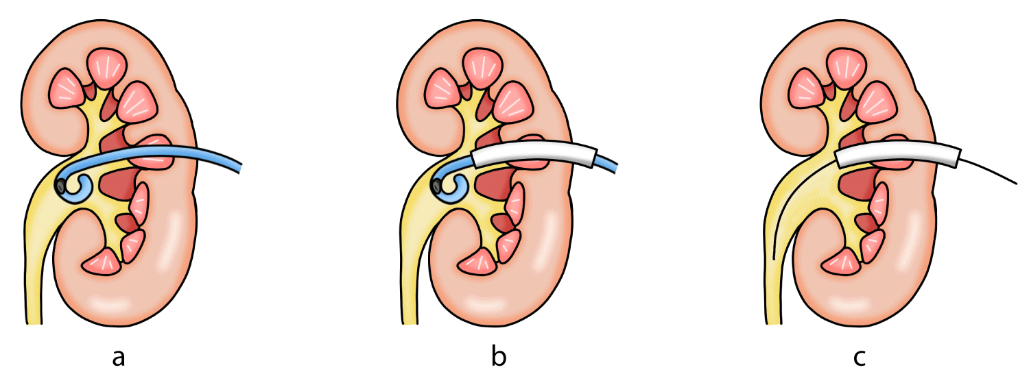 Figure 1a: Schematic diagram showing a pigtail nephrostomy tube with encrustation at the distal catheter.
Figure 1b: Schematic diagram showing the modified vascular sheath advanced over the external segment of the nephrostomy tube and carefully tracked into the renal collecting system under fluoroscopic guidance.
Figure 1c: Schematic diagram of nephrostomy tube exchange using the modified sheath technique. After the modified vascular sheath was positioned appropriately, the indwelling nephrostomy tube was removed while maintaining sheath position. A guidewire was then advanced through the sheath into the collecting system, allowing exchange for a new nephrostomy tube in standard fashion.
View Figure 1
In cases where urine efflux was minimal following removal of the indwelling nephrostomy tube, a 18G or 20G Angio catheter was introduced through the back end of the sheath to occlude the sheath lumen. The needle is then removed and iodinated contrast can be injected through the Angio catheter to fluoroscopically confirm intrarenal positioning prior to guidewire insertion (Figure 2).
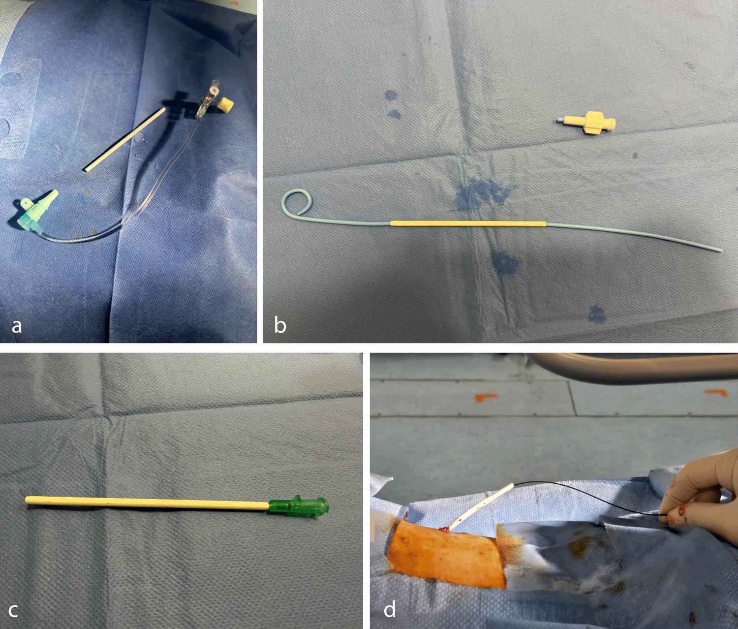 Figure 2a: Clinical photograph of the modified vascular sheath. The stopcock was removed from the vascular sheath to permit advancement over the nephrostomy tube.
Figure 2b: Clinical photograph showing the nephrostomy tube after the hub was cut, following which the modified vascular sheath was advanced over the external segment of the nephrostomy tube.
Figure 2c: An 18G or 20G Angio catheter can be inserted over the backend of the modified vascular sheath, allowing iodinated contrast injection through the Angio catheter to fluoroscopically confirm intrarenal positioning prior to guidewire insertion.
Figure 2d: Clinical photograph showing a guidewire advanced through the sheath into the collecting system. A new nephrostomy tube can then be introduced over the guidewire and positioned in standard fashion.
View Figure 2
In cases where the external length of the indwelling nephrostomy tube was short, or when a relatively long vascular sheath was required, a suture was used to anchor the indwelling catheter. The vascular sheath was then advanced over the catheter using the suture as a stabilizing aid to facilitate controlled advancement into the renal collecting system.
The final study included 20 procedures in 20 patients, with mean age of 76.9. Procedural indications and catheter indwell time are summarized in (Table 1). Technical success rate was achieved in 95% of cases (19 out of 20). In one case, exchange was unsuccessful due to buckling of the vascular access sheath during advancement, which led to loss of secure tract access. Despite attempts at guidewire salvage after removal of the indwelling nephrostomy tube, access to the collecting system could not be re-established, and the procedure was abandoned. The patient was managed conservatively with interval imaging surveillance for recurrent hydronephrosis, with consideration for repeat nephrostomy placement if required. No complications were observed in all 20 procedures.
Table 1: Patients demographics, indications of procedure, catheter indwell time and outcome measurements summarized. View Table 1
Encrustation of indwelling urinary catheters is a well-recognised complication associated with prolonged dwell time, infection, and altered urinary composition, resulting in mineral deposition and biofilm formation along the catheter surface. Although much of the published literature focuses on ureteric stents, the same mechanisms apply to nephrostomy tubes and may lead to catheter fixation within the renal collecting system or tract, rendering routine exchange technically challenging or unsuccessful [1-5].
From an interventional radiology perspective, encrusted nephrostomy tubes pose a distinct procedural challenge. Standard wire-guided exchange techniques may fail, and forceful catheter removal risks bleeding, tract injury, or loss of access. Traditionally described management strategies often involve invasive adjuncts such as tract dilation, endoscopic or lithotripsy-assisted removal, or surgical intervention [1,3]. While effective, these approaches increase procedural complexity, require additional equipment or multidisciplinary involvement, and may not be ideal for patients with significant comorbidities or limited procedural tolerance.
Several minimally invasive techniques aimed at avoiding tract manipulation have been described. Campodonico, et al. reported a non-invasive method for the removal of entrapped self-locking nephrostomy tubes, demonstrating that catheter-based strategies can be effective without escalating to surgical intervention [2]. Farooq, et al. described the use of a vascular sheath to facilitate safe removal of an encrusted nephrostomy tube, highlighting the value of maintaining access to the collecting system while allowing controlled catheter extraction under fluoroscopic guidance [3]. Other reports have described the use of intracorporeal lithotripsy devices, such as lithoclasts, to fragment encrustations; however, these techniques require additional devices and technical expertise [4].
The technique described in this case series is firmly aligned with interventional radiology practice, relying exclusively on fluoroscopic guidance and readily available vascular access equipment. By advancing a vascular sheath over the indwelling nephrostomy tube, access to the renal collecting system is preserved throughout the procedure, allowing safe removal of the encrusted catheter and subsequent tube exchange. Confirmation of intrarenal positioning using urine efflux and contrast injection provides a simple and reproducible safety check prior to guidewire insertion.
Importantly, procedural variations were incorporated to address common real-world challenges. The use of an angiocatheter to occlude the sheath lumen enabled contrast confirmation of collecting system access in cases with poor urine flow, while anchoring the indwelling catheter with a suture facilitated sheath advancement when external catheter length was limited. These adaptations enhance procedural control and broaden the applicability of the technique without increasing invasiveness.
In the present series, technical success was achieved in 19 of 20 cases, demonstrating that this sheath-assisted approach can be reliably performed even when conventional wire-guided exchange fails. The high success rate likely reflects preservation of tract access throughout the procedure and the use of fluoroscopic confirmation prior to guidewire insertion. Importantly, the technique relies on standard interventional radiology equipment and does not require escalation to more invasive adjuncts in most cases.
This study has limitations inherent to its retrospective, single-centre design and the absence of a comparator group. Long-term outcomes were not assessed. Nevertheless, the relatively large number of cases for this uncommon scenario provides meaningful insight into a simple, minimally invasive, and reproducible technique that can be readily adopted by interventional radiologists when standard nephrostomy tube exchange fails.
In conclusion, encrusted nephrostomy tubes represent a challenging scenario in interventional radiology when conventional wire-guided exchange techniques are unsuccessful. The fluoroscopy-guided use of a modified vascular access sheath offers a simple, minimally invasive, and effective method for maintaining collecting system access and facilitating safe nephrostomy tube exchange without the need for tract dilation or endoscopic assistance. This technique may serve as a practical first-line salvage option in routine interventional radiology practice.