
Background: Demodex blepharitis is a highly prevalent chronic ophthalmologic condition that compromises the integrity and proper functioning of the eyelid. There are few statistical data on this condition in patients with type 2 diabetes mellitus (DM2).
Objective: To determine the relationship between Demodex spp. Blepharitis in patients with type 2 diabetes mellitus (DM2) according to their glycemic control.
Methods: Descriptive and cross-sectional study. Fifty-three patients with a previous diagnosis of DM2 who had clinical features of palpebral demodicosis were included-the Demodex spp. The infestation index (InI) was obtained after eyelash biopsy, and a value greater than 0.5 was considered indicative of infestation. Subgroups were formed based on glycemic control (HbA1c < 7% and ≥ 7%).
Results: The study included 424 eyelashes, 106 upper eyelids, 25 women (47.17%), and 28 men (52.83%), with no significant statistical difference between the condition by sex (p = 0.47) or by severity according to the InI (p = 0.73). The median age was 61 years (RIC 57-66), the time of diagnosis of DM2 was 10 years (5-20), and HbA1c was 8.1% (6.5-9.7). The InI was 1.5 (1.2-2.1) overall; the population with uncontrolled diabetes was significantly younger, and the InI was higher in women with adequate glycemic control (p < 0.05). There is no relationship between InI and HbA1c (R = -0.23, p = 0.097) or the time of evolution of DM2 (R = 0.0061; p = 0.097).
Conclusion: The InI by the parasite Demodex spp. does not have a linear correlation with glycemic control or time of evolution (p > 0.05), as one would expect to find, since these are the primary factors associated with established complications in DM2.
Blepharitis, Mite infestations, Type 2 diabetes mellitus, Glycemic control, Cross-sectional studies
DM2: Type 2 Diabetes Mellitus; InI: Infestation Index; HbA1c: Glycosylated hemoglobin
Chronic blepharitis can be caused by the ectoparasite Demodex spp., which has been found in 30% to 60% of cases of chronic blepharitis [1,2]. The mite is present on the skin and its appendages in most humans, establishing a commensal relationship; however, overpopulation can develop, which is known as demodicosis [3].
Demodex spp. has five evolutionary stages, all of which are photosensitive: egg, apodal larva, hexapodal larva (also known as protonymph), octopodal nymph or deutonymph, and adult, which develop after 14.5 days in the host [3].
There are two species that are considered independent: Demodex folliculorum (D. folliculorum) and Demodex brevis (D. brevis). Humans are the only host for both species, which affect the face, ear canal, and eyelids [3,4]. The mite is spindle-shaped with rings on its body. The upper third is called the podosoma, which contains the head with piercing appendages in its mouth that allow it to feed and four short legs on each side, which hinder its motility. The lower two-thirds are called the opisthosoma, which is elongated and narrow at the end, and is longer in the folliculorum species than in the brevis species [3]. Demodex lacks color, hair, and an anus, so digested material remains inside the intestine and explodes or regurgitates fecal matter into the surrounding tissues at the end of its life cycle, causing follicle distension and the characteristic formation of cylindrical scales [3-5].
In ocular demodicosis, D. folliculorum is most prevalent at the base of the eyelashes, where it feeds on skin cells and sebum, leading to anterior blepharitis. D. brevis infests the sebaceous and Meibomian glands, feeding on follicular and glandular epithelial cells, causing posterior blepharitis with Meibomian gland dysfunction, keratoconjunctivitis, and recurrent chalazion [2,5].
The pathogenesis of Demodex is multivariable, obstructing the terminal duct of the Meibomian glands and hair follicles, and therefore granulomatous inflammation, madarosis, trichiasis and dry eye [6] in addition to being a vector for different agents such as Streptococcus, Staphylococcus [4], Bacillus spp. [5], Corynebacterium kroppenstedtii [7], Acinetobacter baumannii, and Klebsiella oxytoca [8].
The prevalence according to gender remains unclear, as different studies reveal a predominance in women [2,9], men [5], while others report no difference between genders [10-11]. Regarding age, most studies report a positive correlation in individuals over 60 years of age [9,10]. Other factors studied include the use of glasses or contact lenses [3], cosmetics, facial creams, low socioeconomic status, poor personal hygiene habits [11], and systemic conditions such as immunosuppression [3,7], metabolic syndrome [11,12], skin diseases including rosacea and seborrheic dermatitis [3,4], gestational diabetes [13], and type 2 diabetes mellitus (DM2) affecting the ocular [14,15] and facial areas [16,17]. In addition, a relationship has been established between ocular demodicosis and facial dermatosis [18,19], specifically with rosacea [20], while the influence of treatment between these entities has also been determined [21].
Currently, studies have correlated glycemic control with the presence of cutaneous [12,17] and eyelid [10,22] demodicosis. Therefore, the purpose of this study was to determine the infestation index (InI) in patients with type 2 diabetes mellitus (DM2) and to elucidate whether there is a relationship with glycemic control.
This study followed the principles of the Declaration of Helsinki; it was submitted for evaluation and complied with the requirements of the Ethics and Research Committee of the Mexican Social Security Institute under institutional registration number R-2022-2108-047. The interventions performed on the study subjects were obtained with prior signed informed consent.
This cross-sectional study was conducted between November 2022 and June 2023 at the General Hospital of Zone No. 20, La Margarita, of the Mexican Institute of Social Security. Fifty-three patients with DM2, of both sexes, over 18 years of age, with clinical characteristics of blepharitis caused by Demodex spp. participated. The exclusion criteria were as follows: known cause of immunosuppression, history of contact lens use, ophthalmic surgery within the last 6 months, pregnancy, breastfeeding, and patients who had recently received topical or systemic acaricides. All participants underwent an interview, ophthalmological examination, and laboratory tests.
Under a slit lamp, four eyelashes were selected from each upper eyelid, one from the temporal portion, one from the nasal portion, and two from the medial portion (modified Costón method), which had cylindrical scales or a brittle appearance [4,23]. Using fine tweezers, the eyelash was rotated clockwise and counterclockwise before removal [23]. The eyelashes were placed on a slide, a drop of alcohol was added to dissolve the scales and allow the parasite to be observed properly, and they were covered with a cover slip and observed under a microscope at 10X-400X magnification.
The diagnosis of infestation was made by obtaining the InI for Demodex spp., which is obtained by dividing the total number of specimens in any evolutionary stage observed by the total number of eyelashes removed. In the present study, only the nymph and adult evolutionary stages of any species were included, the main reason being the initial difficulty in identifying the larval and egg stages. An index value greater than 0.5 was considered an infestation [4,24].
The diagnostic criteria of the American Diabetes Association 2022 were used for the diagnosis of DM2. The duration of DM was divided into years and grouped into two categories: < 10 years and ≥ 10 years. Glycemic control was determined by glycosylated hemoglobin (HbA1c) and classified as controlled with HbA1c < 7% and uncontrolled with HbA1c ≥ 7% [25].
The data were analyzed using the free statistical software R. Descriptive statistics are reported in frequency and percentage for qualitative variables, and for quantitative variables with abnormal distribution, median (ME) and interquartile range (IQR) were used. Qualitative variables were recorded using bounded catalogs.
The Mann-Whitney U test was used to demonstrate differences between groups with quantitative variables. Spearman's correlation coefficient, a non-parametric test, was used to determine the correlation between the main variables being studied, with a significance level of α < 0.05 to ensure 95% confidence.
Fifty-three patients diagnosed with DM2, and blepharitis caused by Demodex spp. were analyzed. Twenty-five (47.17%) women and twenty-eight (52.83%) men were included, with no statistically significant difference in conditions by sex (p = 0.47); the median age was 61 (57-66) years.
A total of 424 eyelashes from 106 upper eyelids were examined, accounting for a total of 723 Demodex spp. and a median of 10 (11-17) parasites in the hyphal (14%) or adult (86%) stage of development (Figure 1). All patients were infested with D. folliculorum, and only 12 (22.64%) had D. brevis, all of which were in the adult stage, with 8 (15.09%) female patients and 4 (7.54%) male patients. The ratio between D. brevis and D. folliculorum was 1:35, and only 46 (87%) of the 53 patients reported symptoms (Table 1, Figure 1).
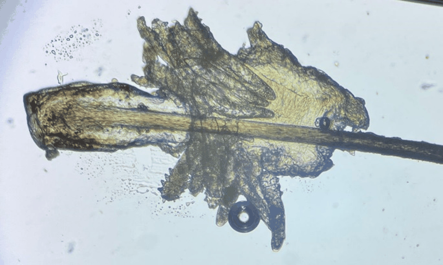 Figure 1: Infestation by Demodex spp.
*The hair follicle of an eyelash with multiple parasites surrounded by cylindrical scales is observed.
View Figure 1
Table 1: Sociodemographic and clinical characteristics of the population. View Table 1
The median time since diagnosis of DM2 was 10 (5-20) years, of which 27 (51%) had been diagnosed within the last 10 years and 26 (49%) more than 10 years ago.
The median HbA1c was 8.1 (6.5-9.7); 18 (34%) patients had adequate glycemic control with an HbA1c < 7% (male:female 6:12), while the rest were uncontrolled with an HbA1c ≥ 7% (M:F 22:13).
The median InI was 1.5 (1.2-2.1) overall, 1.38 (1.25-2.63) for females, and 1.56 (1.25-2.03) for males, with no statistically significant difference between sexes (p = 0.73). The InI is significantly higher in patients aged 61 years or older [< 61 years = 1.3 (1.1-1.9) vs. ≥ 61 years = 1.75 (1.2-2.7), p = 0.04] (Table 2).
Table 2: Comparison and relationship of the InI in the population. View Table 2
The InI according to the time of evolution is 1.78 (1.25-2.34) for the group with less than 10 years of diagnosis and 1.64 (1.25-2.0) for the group with more than 10 years, with no statistical differences (p = 0.55).
When comparing the HbA1c < 7% and HbA1c ≥ 7% groups, a statistical difference was found in age [Control: Uncontrolled 65 (60.75-71.75) vs. 59 (55.5-62.5) p = 0.005], with the uncontrolled group being younger (Table 3).
Table 3: Comparison according to glycemic control. View Table 3
There were no differences in the InI of the groups [C: D 1.75 (1.25-2.84) vs. 1.37 (1.19-1.94) p = 0.12], nor between males according to glycemic control [C: D 1.25(1.25-1.63) vs 1.69 (1.25-2.09), p = 0.46]; however, there was a difference in females [C:D 2.69(1.25-3) vs 1.25(1.13-1.38), p = 0.02]. No differences were found in the time of evolution [C: D 10(5-20) vs 10(6-15.5) p = 0.63]; as well as in the count of D. folliculorum [C:D 14 (10-22) vs. 11(9-15) p = 0.16] and D. brevis [C:D 0(0-1) vs. 0(0-0) p = 0.06].
When examining the correlation between InI and the variables, a significant correlation was found with the degree of cylindrical dandruff (R = 0.36, p = 0.008). In contrast, no correlation was found with HbA1c (R = -0.23, p = 0.097) or any other variable (Figure 2).
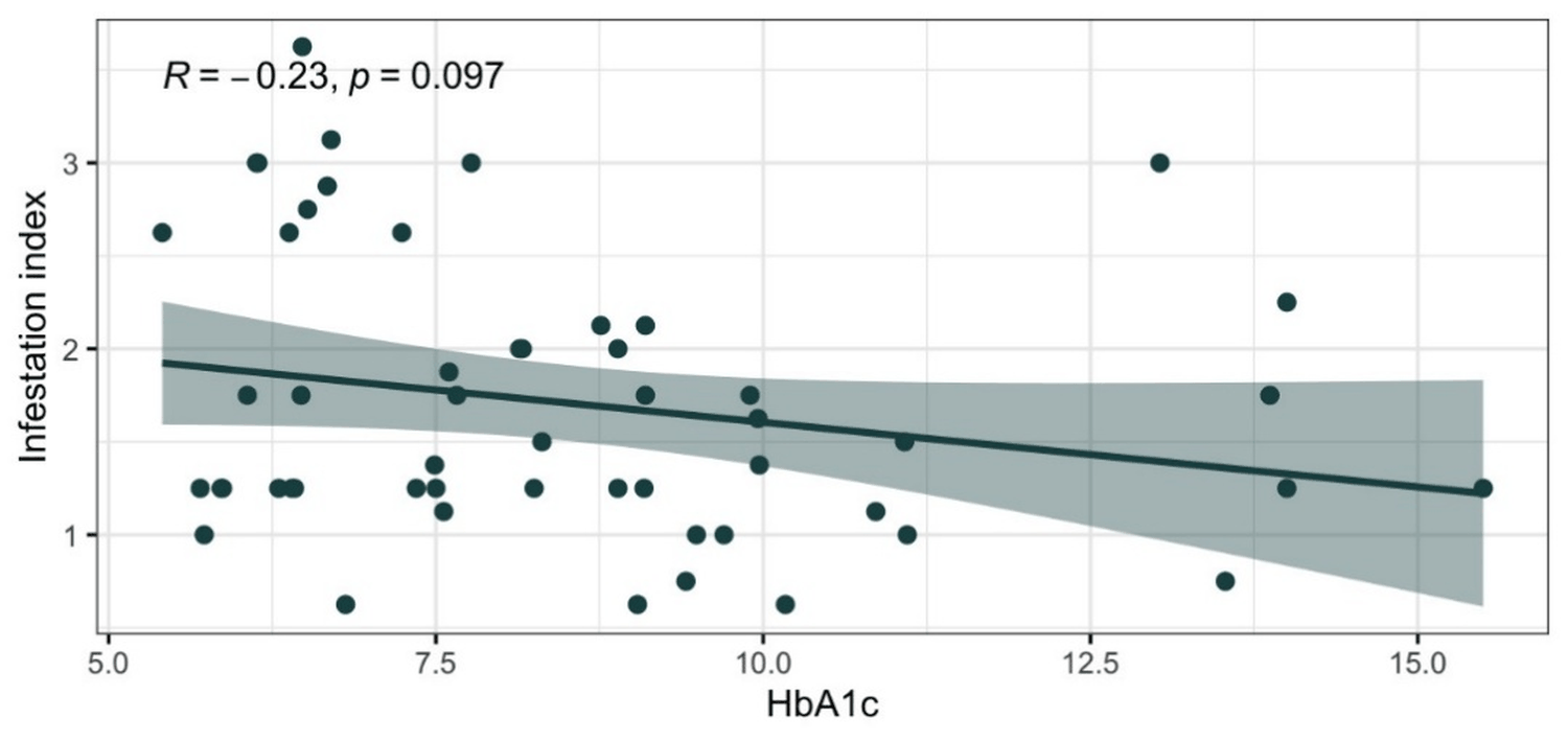 Figure 2: Scatter plot of the relationship between glycated hemoglobin and the Demodex spp. infestation index.
The shaded area corresponds to the confidence interval of the trend line.
View Figure 2
Chronic blepharitis, as in the case of eyelid demodicosis, is a highly prevalent ophthalmological condition that affects the integrity of the eyelid and its proper function, which has an impact on quality of life [26,27].
Several studies correlate demodicosis with DM2. Cutaneous demodicosis is more prevalent and easier to diagnose because a standardized method and established criteria are available [17,19]. This is not the case with eyelid demodicosis, as some studies only consider the presence or absence of the parasite [2,11]. Some use the InI, [4,24] and others use different diagnostic criteria, which vary even within the same country, such as Chang Huang, et al. (2021, China) [10], who used criteria based on the 2018 consensus of Chinese experts, and Zhang N, et al. (2023, China) [22], who used their own criteria. However, the association between the entities is positive in both studies.
In the population studied, the median age of 61 years (57-66) defines the statistically significant difference in InI [< 61 years = 1.3 (1.1-1.9) vs. ≥ 61 years 1.75 (1.2-2.7), p = 0.04], in agreement with López-Ponce D, et al. (2017) [4], who mention a higher prevalence of the parasite with respect to age; and with Mongi F, et al. (2018) [24], who found significant associations in patients over 60 years of age (p < 0.001).
There is no preference for gender in demodicosis in the present study (p = 0.47), which is consistent with most previous studies [10,11].
The most common parasite found was Demodex folliculorum, with a Brevis/Folliculorum ratio of 1/35, which is consistent with the study by Lingyi Liang, et al. (2021, China) (1/24) in the general population and with Zhang N, et al. (2023, China) [22] (1/3) in the population with DM2.
As mentioned by Mongi F, et al. (2018, Argentina) [24], although some patients may remain asymptomatic, most report symptoms with an InI > 0.5; this data was corroborated, as up to 87% of patients reported symptoms, the main one being eyelid itching; this contrasts with Lingyi Liang, et al. (2021, China), who report eye fatigue and photophobia as the predominant symptoms for this age group.
The median number of years since diagnosis of DM2 is 10 (5-20) years, as is the control with HbA1c of 8.1% (6.5-9.7), which are risk factors for developing microvascular, macrovascular, and immunological complications that could be related to the presence of the parasite.
In terms of glycemic control, the proportion of patients is inversely proportional to the results obtained by Zhang N, et al. (2023, China) [22]; (C:D = 34%:66% vs. 66%:34%), suggesting greater glycemic uncontrolledness in our population. However, we agree that patients with poor control are significantly younger [C: D= (66.5 ± 6 8.4 vs 64.2 ± 6 8.6 years, p = 0.02), and (66 ± 8.87 vs 59 ± 6.39, p = 0.005)] [22].
Although there is no significant difference according to sex in the InI [M: H = 1.38(1.25-2.63) vs 1.56 (1.25-2.03), p = 0.47], there was a difference according to glycemic control in females (C:D 2.69 vs. 1.25, p = 0.002), being higher in controlled patients, which could be explained by the age difference between the two groups.
No statistical differences were found in the InI based on glycemic control [C:D 1.75 (1.25-2.84) vs. 1.37 (1.19-1.94); p = 0.12], as reported by Zhang N, et al. (2023, China) [22] with the count [C:D = 3 (0-11.9) vs. 5 (0-14.6); p = 0.02], nor the count according to species. However, we both agree that there is no difference in the subgroups according to the time of evolution [OR 1.01 (0.98, 1.04), p = 0.5] and [C: D 10 (5-20) vs 10 (6-15.5), p = 0.63]. There was also no difference in the InI according to years of diagnosis [< 10 years: ≥ 10 years = 1.78 (1.25-2.34) vs 1.64 (1.25-2.0); p = 0.55] [22].
The results differ from those reported by Chang Huang, et al. (2021, China) [10] in the linear correlation between Demodex spp. infestation and population age (R = 0.24; p = 0.088 vs R = 0.299, p < 0.001) as they show little significance, as well as the time of evolution of the underlying disease (R: 0.0061; p = 0.097 vs R: 0.264, p < 0.001).
The semi-quantitative estimation of cylindrical dandruff was performed according to López-Ponce, et al. (2017, Chile) [4], and a positive correlation was observed between the amount of cylindrical dandruff and the InI (R = 0.36; p = 0.008).
Regarding the main objective of this study, we did not find a linear correlation between InI and HbA1c (R = -0.23, p = 0.097).
Among the limitations of this study are the lack of standardized diagnostic criteria, its single-center nature, and the sample size, which was based on statistics reported in the medical unit. These limitations should be considered in future studies.
The findings of this study are relevant, as it is the first state, and nation-wide study to describe the rate of Demodex spp. infestation in patients with type 2 diabetes mellitus.
The severity of Demodex infestation does not correlate with glycemic control or disease duration, as would be expected, given that these factors are the primary triggers of established complications in diabetes. However, infestation does affect quality of life due to eyelid inflammation, Meibomian gland dysfunction, and consequently dry eye, as has been established in previous studies.
Conceptualization: Tiffany Garcia, Elvira Cantu. Methodology: Tiffany Garcia, Luis Gomez, Daniel Canaan. Data creation: Tiffany Garcia. Formal analysis: Tiffany Garcia, Luis Gomez. Investigation: Tiffany Garcia. Original draft writing: Tiffany Garcia, Luis Gomez. Writing, revision, and editing: Tiffany Garcia, Luis Gomez. Approval of the final version of the manuscript: all authors.
Authors declare no conflict of interest.
Nil.
The authors would like to thank the Ophthalmology Department of Zone 20 General Hospital and the CADIMSS Department of UMF 55 of the Mexican Social Security Institute for their support in providing the material and human resources necessary to carry out this project. As well as to Elaine Fernanda García Lujano for her support in writing.