
TLE: Transvenous lead extraction
SVC: Superior Vena Cava
BB: BridgeTM Balloon
Introduction: In the case of an injury to the superior vena cava (SVC) during transvenous lead extraction (TLE), immediate sternotomy is usually necessary. The use of a rescue balloon, which tamponades temporally the SVC tear, has shown to be associated with an improved outcome in these situations. We aimed to test a prophylactic endovascular placement of the rescue balloon during TLE to facilitate placement in case of emergency.
Methods: 10 consecutive patients undergoing TLE were enrolled for this case series. The rescue balloon was test-inflated under fluoroscopic guidance in all cases, staged during the extraction procedure, and re-inflated without fluoroscopic guidance at the end of the extraction.
Results: Mean age of the population was 68±17 years with a mean LVEF of 43.3±14.0%. The main indication for TLE was lead dysfunction (80%). The balloon was successfully inflated in all cases and staged either outside the body (1 case), in the SVC (1 patient), in the IVC (3 patients) and in 5 patients within the introducer sheath. In 90% of the patients, the balloon could be re-inflated without the use of fluoroscopy at the end of the procedure. Mean reinflation time was 43±11seconds after a mean balloon dwell time of 20.5±11.2 minutes. There were no clots identified on the balloon surface.
Conclusion: The prophylactic use of a rescue balloon during transvenous lead extraction procedures is safe. In case of complex interventions, it can be staged intravascularly without the risk of clot formation if the dwell time is minimized.
Transvenous lead extraction, Occlusion balloon, Superior vena cava rupture
Transvenous lead extraction (TLE) for device infection or lead malfunction is increasingly being performed [1]. Even though the risk for major complications is relatively low in experienced centers (1.7 % according to the results from the ELECTRa registry) [2], some complications may have devastating outcomes and complication rates may be higher in developing lead extraction centers [3]. Injury to the venous wall during TLE, especially at the level of the superior vena cava (SVC), is often unpredictable and the most feared event due to the high mortality rate despite immediate surgical repair [4-6]. To improve management and finally survival of this complication, an endovascular occlusion balloon (Bridge TM , Philips, Colorado Springs, CO, USA) was developed [7-10]. The Bridge TM balloon is a compliant, over-the-wire balloon that can be advanced percutaneously from a femoral venous access to the level of the SVC offering a temporary occlusion in case of SVC laceration until surgical repair is performed [7]. In a first retrospective cohort analysis of 35 patients suffering from an SVC injury during TLE, the survival rate was 100% when the Bridge TM balloon (BB) was used compared to 50% when the rescue balloon was not available [11]. A follow-up analysis of 116 SVC events confirmed a significantly higher survival rate if the Bridge TM balloon was used (88.2% versus 56.9%) [7]. However, the use of such a percutaneous balloon may come with potential side effects. Clot formation on the surface of the balloon has been described, especially when the device is staged intravascularly for an extended period of time during TLE and, as usual, no heparinisation is performed during the intervention to prevent excessive blood loss [12,13]. A Food and Drug Administration’s Manufacturer and User Facility Device Experience (MAUDE) database analysis found thrombotic clot formation on the balloon surface in 18% of reported adverse events but only in 2% a significant pulmonary embolism was reported [13]. Other potential adverse events described with the balloon were a compromised balloon integrity (either seen as air/blood aspiration, a tear on the balloon surface or unexpected rupture of the balloon) or an inability to advance the necessary stiff wire through the balloon catheter inner lumen [13]. Despite these above-mentioned risks, especially considering time-dependent clot formation, the balloon can be staged prophylactically at the level of the inferior vena cava or within the sheath in high-risk interventions to avoid a placement delay in case of an SVC tear [7,9]. In a prospective cohort analysis the risk of clot formation was estimated as high as 67% if the balloon was staged intravascularly for the entire procedure (dwell time 128±74 min), but only 4.8% if the exposure time of the Bridge TM balloon was limited to the extraction intervention itself only (dwell time 25±18 min), highlighting the time-dependent risk of thrombosis [12]. However, placing an intravascularly staged Bridge TM balloon at the place of SVC laceration still requires time and fluoroscopy, both of which may be limited in availability during an acute SVC complication. This case series of 10 consecutive patients referred for TLE at our center was performed to investigate whether a standardized, abbreviated protocol for the deployment of the Bridge TM balloon is safe, but also whether it is possible to restage the balloon at the level of the SVC without fluoroscopy.
10 consecutive patients undergoing TLE at the University Heart Center Zurich, Switzerland, were enrolled for this case series. All patients have given written informed consent. Patient demographic and clinical baseline characteristics, device as well as lead properties and TLE procedure related parameters were collected. Data collection was performed trough studying of operation reports, discharge reports and chest x-ray. Continuous variables are expressed as mean ± standard deviation, while categorical variables are expressed as absolute numbers and percentages.
All interventions were performed under general anaesthesia in a hybrid operating room and performed by 2 experienced electrophysiologists. Venous access via the internal jugular vein as well as bifemoral access was acquired in all subjects. Invasive blood pressure monitoring was performed via radial arterial catheter. The choice of an additional femoral arterial access was left to the discretion of the operator. A superior approach via the implant-related vein was always the primary TLE method unless this approach had previously failed. If simple traction failed, the leads were prepared using a lead locking stylet (Liberator®, Cook Medical, Bloomington, IN, USA) and lead insulation secured using a compression coil (One-Tie®, Cook Medical, Bloomington, IN, USA). High-voltage conductors were not separately secured. All leads were extracted by simple traction or, if necessary, by using mechanical rotational tools. A cardiothoracic surgeon was informed and immediately available in the event of a complication. All patients were admitted overnight to the intermediate care unit until the next morning. Anticoagulation with Non-Vitamin K-oral anticoagulants (NOACs) was interrupted >48h prior to the intervention, while the use of vitamin K antagonists was adjusted to achieve an international normalized ratio (INR) of around 2.0 prior to the procedure. Oral anticoagulation was reinitiated 48 hours after intervention.
Ultrasound-guided, femoral venous access via the right femoral vein was obtained in all cases. A standard J-wire (0.035”) was inserted and a short 9Fr sheath introduced. The J-wire was then replaced with a 0.035” Amplatzer Super-Stiff TM wire (Boston Scientific) which was advanced up to the level of the right internal jugular, the subclavian or brachiocephalic vein. The 9Fr sheath was then replaced with a 30-45cm 12Fr sheath (Cook Medical). Next, the device pocket, device and the leads were prepared as described above. As soon as the leads were ready to be extracted, the Bridge TM balloon was advanced to the SVC and (test-)inflated. For inflation a 60cc mixture of contrast media and saline (12cc contrast media, 48cc saline) was used and the volume needed for appropriate inflation was documented. Fluoroscopy images were taken at that position in different angulations (RAO 30°, AP, and LAO 40°) and the level of the balloon catheter position relative to the 12Fr sheath was marked. Then, the balloon was deflated and staged either intravascularly, inside the sheath or outside the body. After the TLE procedure was successfully completed, the balloon was readvanced without fluoroscopy up to the previous marking on the balloon catheter. If this was successful without any resistance, the balloon was re-inflated using the same volume as during test inflation and the appropriate correct position confirmed then by fluoroscopy. During the entire procedure and after removal, the balloon surface was checked for clot formation. A relevant clot formation was defined as a thrombus size > 10 mm [12]. Additionally, the balloon was checked for surface damage. Balloon handling including potential other side effects apart from clot formation (injury to the vessel wall, dislodgement, air embolism, change in hemodynamics during test inflation) was documented.
Continuous variables were compared using paired t-test. Categorical variables were compared using the v2 test or Fisher’s exact test as appropriate. A two-tailed a level of 0.05 was used to define statistical significance. All analyses were performed using SPSS (SPSS, V 24.0, IBM Corp., Armonk, NY, USA).
Baseline patient data are summarized in Table 1. All patients were male with a mean age of 68±17 years and with a mean LVEF of 43.3±14.0%. Half of the population had atrial fibrillation in their medical history and were on anticoagulation using a non-vitamin K oral anticoagulant. The main indication for TLE was lead dysfunction (80%). Single coil defibrillator leads were present in 50% of the cohort and coronary sinus electrodes in 30%. The mean number of leads extracted was 1.7±0.8 with a mean dwell time of 6.3±4.2 years (Table 2); the oldest lead was implanted 14 years prior to TLE. A mechanical rotational tool (either Cook TM Evolution or Philips TM TightRail) was used in 70 % of TLE procedures, while in 3 patients the leads could be extracted with traction only. The mean intervention duration was 113.8±42.1. minutes. All targeted leads were successfully extracted via a superior approach without femoral snaring performed during TLE.
Table 1: Baseline clinical characteristics. View Table 1
Table 2: Lead extraction parameters. View Table 2
In all patients, a 12Fr sheath was inserted from the right femoral groin and a super stiff wire was advanced to the level of the right internal jugular vein. Next, the pocket and the leads were prepared for the extraction procedure. Just prior to the lead extraction procedure itself, the balloon was placed at the level of the SVC in all patients for a test-inflation and fluoroscopy images taken in 3 angulations (AP, RAO 15-20°, LAO 30-40°; Figure 1). After full inflation and confirmation of a stable balloon position, the level of the Bridge TM balloon catheter position relative to the 12Fr femoral sheath was marked and the balloon deflated. The balloon was staged outside the vasculature in 1 case, in the SVC in 1 case, in 3 patients at the level of the IVC, and in 5 patients in the long 12Fr sheath (Table 3). There was no need to inflate the balloon during the TLE due to emergency reasons in any patient. After completion of the lead extraction intervention itself, the balloon was re-inserted blindly without the use of fluoroscopy and inflated again using only the physical markers of the marked lumen and noted filling volume. (radiation was only used for confirmation of correct position) using the same amount of contrast/saline mixture as during the initial inflation. The Bridge balloon’s position was then checked against its original placement position.
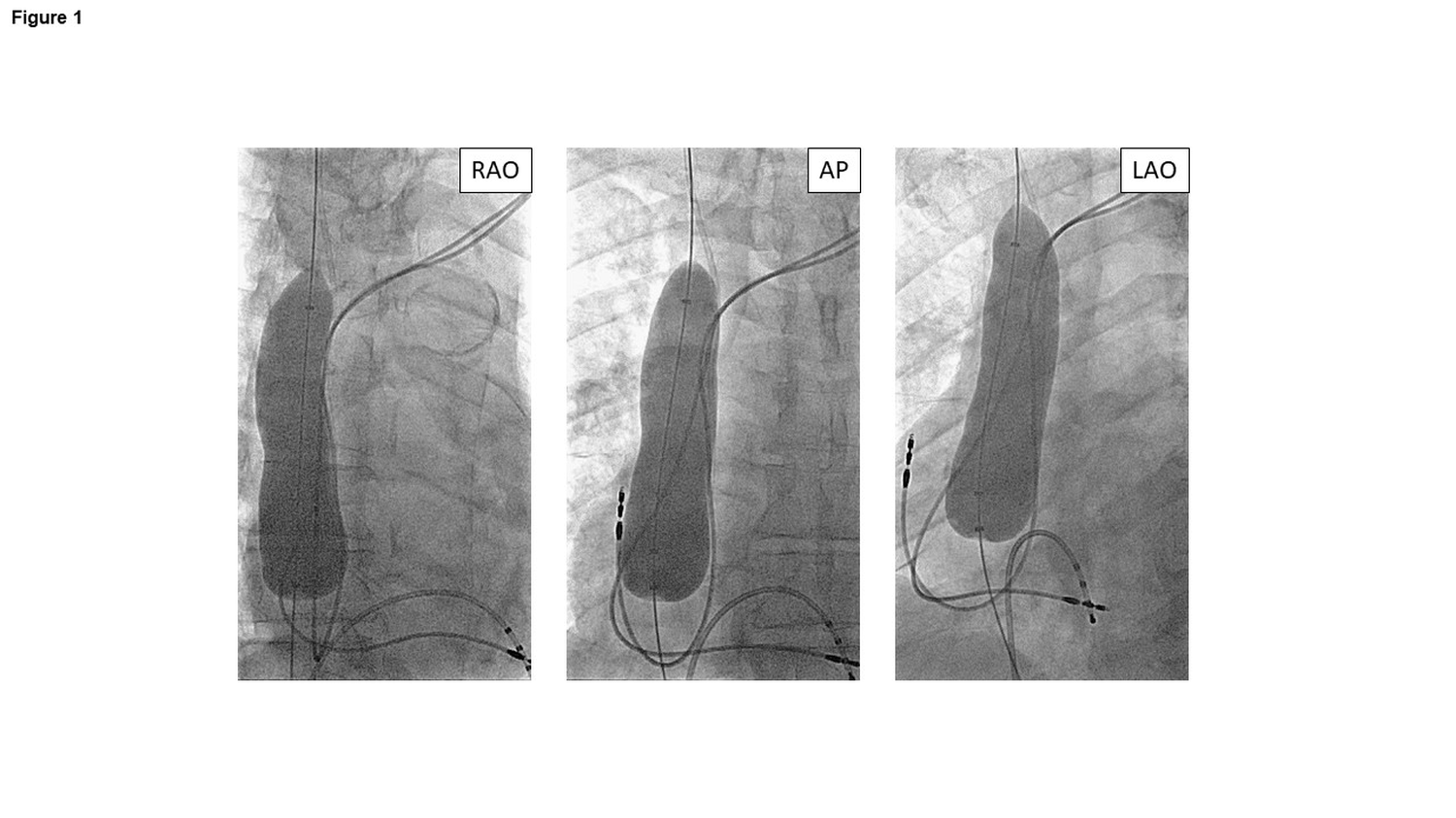 Figure 1: Inflated BridgeTM balloon in different projections (RAO, AP, LAO).
View Figure 1
Figure 1: Inflated BridgeTM balloon in different projections (RAO, AP, LAO).
View Figure 1
Table 3: Peri-interventional parameters of BridgeTM balloon inflation. View Table 3
In 90% of the patients (9/10), reinflation without fluoroscopy resulted in a successful inflation in the targeted area of the SVC (Table 3). In one patient, balloon inflation was too inferior within the SVC and as a consequence dislodged into the right atrium as visualised by fluoroscopy; however no further sequel resulted from the dislodgement. Mean re-inflation time was 43±11seconds. Mean dwell time of the balloon was in total 20.5±11.2 minutes. Clot formation (investigated either via transesophageal echocardiography or via visual inspection on the balloon surface after removal) was not observed in these 10 cases. No major complications occurred during initial placement of the Bridge TM balloon or during re-positioning. However, in one patient suffering from severe heart failure (with a LVEF < 15%), the inflation of the Bridge TM balloon resulted in a significant transient reduction of cardiac output due to preload reduction during the time of the Bridge TM balloon inflation, from which the patient fully recovered after deflation.
TLE nowadays is an increasingly performed intervention with a low major complication risk as well as a periprocedural mortality rate below 1% [2]. The most feared complication, albeit not very frequent, is laceration of the SVC which is associated with a high mortality rate of nearly 50 % [2,4-6,11]. While injury to the venous vessel wall can theoretically occur at any position, in a fifth of the patients the tear is at the level of the SVC but above the pericardial cavity resulting in exsanguination of blood into the pleural space, and in a third it is at the level of the junction between the SVC and the right atrium [5]. The latter may cause both pericardial tamponade as well as haemothorax. An SVC tear of ³ 2 cm in size will lead to complete exsanguination within 10 minutes and to a mortality rate of more than 30 % despite surgical intervention [10]. Hence, immediate recognition of such a complication is essential to improve outcome as well as rapid, at least temporary, occlusion of the laceration site to reduce exsanguination to offer time for the extraction team to prepare for surgical repair [7,8].
The Bridge TM occlusion balloon is an 80-mm compliant balloon designed to tamponade the site of injury in case of an SVC tear. A first retrospective analysis demonstrated that all patients with an SVC tear and Bridge TM balloon deployment survived, while the mortality rate was 50 % in the 26 cases where the Bridge TM balloon was not used 11 . These data are supported by a recent analysis of 116 confirmed SVC tear cases where the use of the Bridge TM balloon was associated with a survival rate of 88.2 % in comparison to 56.9% if the balloon was not used or not properly used [7]. This underscores that both a fast and correct deployment of the balloon is essential for a beneficial outcome since any relevant delay or inappropriate deployment will result in a significant blood loss [7,9]. There is therefore a minimum standard set-up from the recent Best Practice Protocol [7] (Figure 2), which recommends that in all patients undergoing TLE, a 0.035” stiff guidewire is advanced from the femoral vein through the SVC to any internal jugular, subclavian or brachiocephalic vein. Further, a 12Fr introducer sheath must be inserted over the 0.035” stiff wire and a 60cc prefilled syringe (12cc contrast media, 48cc saline) for balloon inflation is ready to use. In patients at increased risk for vascular injury during TLE, such as the presence of dual coil ICD leads, long lead dwell time, low patient’s BMI, female gender, chronic kidney disease but also low operator volume/experience, intravascular staging of the Bridge TM balloon preferentially at the level of the IVC or within the introducer sheath can be considered including test deployment to record to necessary inflation volume and the correct position of the balloon [14,15]. Indeed, a prophylactic positioning of the balloon intravascularly significantly reduced the time to correctly deploy the balloon in case of an SVC tear [9]. If the balloon is, in addition to the minimal requirements mentioned above, staged prophylactically at the level of the IVC, time to correct deployment was <15 seconds according to this multicentre analysis as compared to 120 seconds, if the balloon had to be unpacked in case of an emergent use [9]. Furthermore, it is potentially also more challenging to correctly position the balloon in case of an emergency.
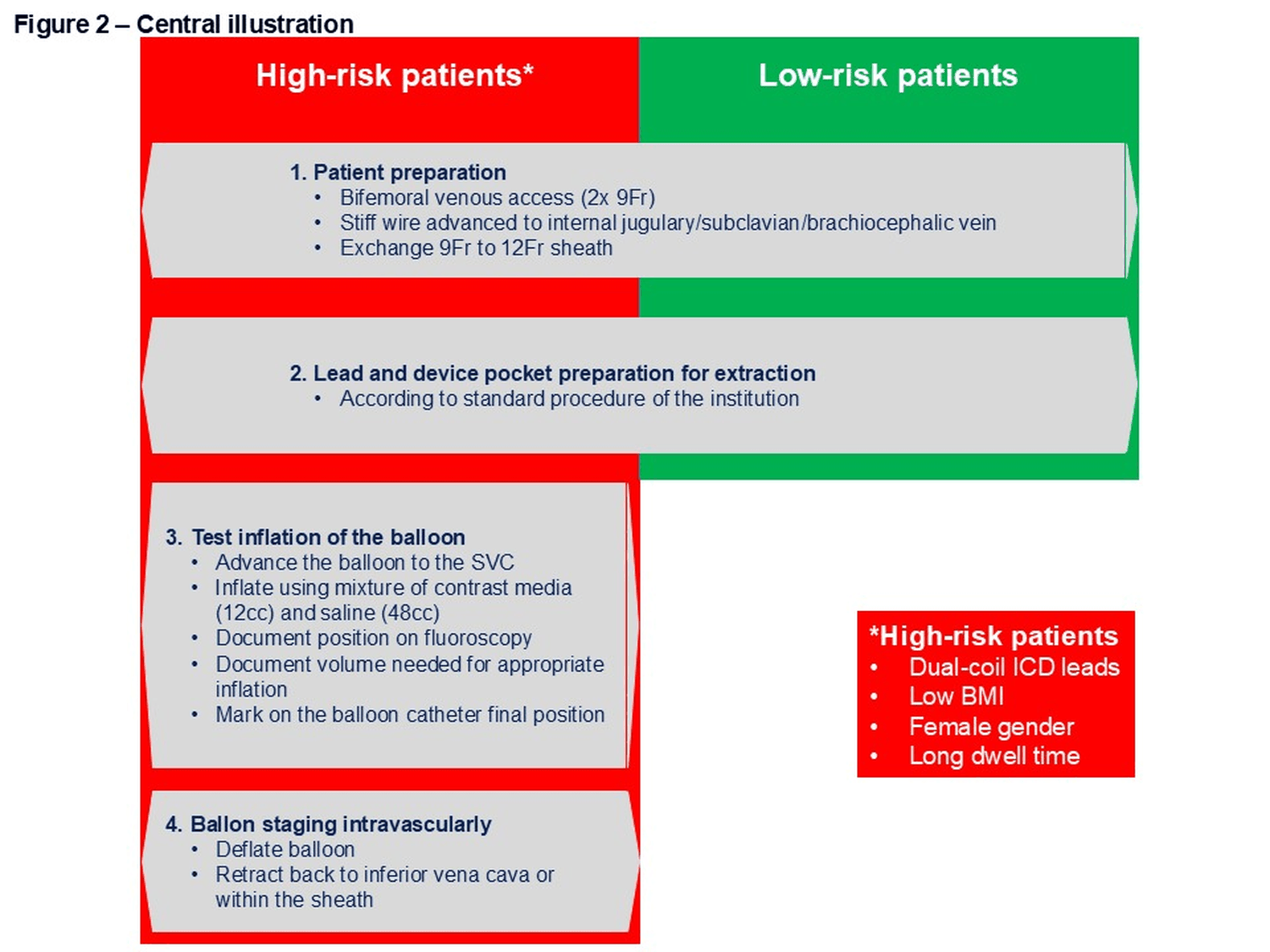 Figure 2: (Central illustration). Recommendation for the use of the occlusion balloon during transvenous lead extraction.
View Figure 2
Figure 2: (Central illustration). Recommendation for the use of the occlusion balloon during transvenous lead extraction.
View Figure 2
Even though it has been documented that prophylactic staging of the Bridge TM balloon was not associated with an increased risk of pulmonary embolism or deep vein thrombosis 9 , there is some recent concern of clot formation on the surface of the balloon. Pre-clinical in vitro and in vivo studies have evaluated the surface thrombogenicity showing the balloon caused some activation of the intrinsic coagulation pathway [9] and that microclot formation was observed in the swine vasculature if the balloon was deployed for 45 minutes 10 . The clinical consequences in humans however are less clear. Pothineni, et al., [12] has shown a high incidence of balloon-related thrombus formation (66.7%) if the balloon dwell time is long (128±74 min), compared to 4.8 % if the intravascular dwell time of the balloon is reduced to 25±18 minutes. Likewise, a Food and Drug Administration’s Manufacturer and User Facility Device Experience (MAUDE) database analysis described clot formation in 17 cases (18%) on the balloon surface [13]. In our cohort of 10 subjects and a balloon dwell time of 20±10 minutes, no clot formation (predefined with a size of > 10mm) was seen either during the intervention or by direct visual inspection after removal of the balloon, which is strongly in line with the finding from Pothineni, et al., [12]. Interestingly, even in the presence of clots with a mean length of 3.99±1.4 cm in the long dwell time group, only 1 out of 14 patients with a clot formation developed a pulmonary embolism (on day 3 after the intervention) for which he had been started on oral anticoagulation [12]. In line, another report from 21 patients where the balloon was placed prophylactically at the level of the IVC or SVC, no thromboembolic complications were observed over a follow-up period of 6.8±3.7 months [9]. Taken together, the risk of clot formation on the surface of the Bridge TM balloon is not zero, but usually clinically not relevant if the dwell time is kept as short as possible and therefore the Bridge TM balloon is only staged intravascularly during the TLE procedure itself rather than the entire transvenous device intervention.
The Bridge TM balloon can only work properly and be of benefit if it is deployed correctly; and there are some aspects in the handling of this tool which are essential. The 0.035” stiff wire must be advanced far enough to the level of the internal jugular, subclavian or brachiocephalic vein to ensure adequate support at the level of the SVC. Indeed, a relevant reason for malfunction of the balloon was an unintentional removal/displacement of the stiff wire before balloon deployment preventing therefore a good enough stability during inflation [7]. Furthermore, equally important to the correct position of the wire is the correct access preparation using a 12Fr sheath in all cases [7]. In emergency, hypotonic shock conditions it may not be possible to exchange a smaller diameter sheath with the necessary 12Fr sheath as has been observed in the past and therefore, all operators performing TLE are advised to insert a 12Fr sheath from beginning of the intervention 7 . If an operator decides to stage the Bridge TM balloon prophylactically, it is recommended to test-inflate the device to record the volume necessary to inflate the balloon and to mark the position on the catheter as a guide for how far to advance the balloon again in case of an SVC tear. The SVC usually has an average length of 6 cm with half of the SVC being outside the pericardial space [16]. Obviously, the balloon must be inflated and tamponade the vessel at the level of the SVC tear. In this context, one need to keep in mind that in cases of a rather inferior SVC tear, balloon position may not be as stable as necessary. Indeed, we have observed in one patient that the Bridge TM balloon was deployed within the SVC but close to the junction to the right atrium, and consequently dislodged into the right atrium after inflation. This happened most likely as a combined result from the pushback due to the venous blood flow while insufficient anchoring of the balloon on the venous vessel wall was present, and the pulsatile contractility of the beating heart. It may be necessary to place the balloon rather inferior within the SVC if an injury occurs (or is being suspected to have occurred) at the inferior level of the SVC; however, in these situations we recommend a special attention to the stability of the balloon once inflated. In general, the compliance of the balloon allows a safe inflation independently of the presence of electrodes or a venous side branch or even an SVC stenosis [8].
Inflation of the balloon at the SVC for half an hour is safe and does not result in a physiological or neurological compromise as shown in a swine model [10]. This may be different in patients with special cardiac conditions. Indeed, in one patient of this cohort suffering from severely impaired left ventricular function with an LVEF on echocardiogram of 15 %, we have observed a significant drop in blood pressure after balloon inflation which most likely resulted from the impaired venous backflow and hence a reduced preload necessary for cardiac function. Indeed, acute occlusion of caval veins have been shown to be associated with a decrease in cardiac output and a reduction in mean arterial blood pressure in heart failure patients. In the case series of Pothineni, et al., the mean LVEF was 38.5±13.9% and hence also significantly reduced, but a hemodynamic relevance was not described [12]. This may be an underreporting bias; however, since in all cases performed in this institution, bifemoral venous access was acquired at the beginning of the procedure, intravascular fluid balance as well as application of inotropic medication most likely could be managed via the contralateral femoral venous access bypassing the blocked venous blood flow from the upper part of the body during inflation of the balloon. This is an aspect of clinical relevance: In addition to the minimal standard which includes placement of a stiff wire at the correct place and having a 12Fr sheath inserted from beginning on the TLE [7], we strongly recommend to have always a second, contralateral femoral venous line in situ to have a secured and large access in case of an injury at the SVC level, which guarantees access for fluid and emergency medication.
None.
This study was funded by Philips, Colorado Springs, CO, USA
The gathering and analysis of all patient data for this paper adheres to the Swiss Ordinance on Human Research with the Exception of Clinical Trials and was approved by the local ethics committee (BASEC-NR: 2018-01540). All patient data was anonymized.
Written informed consent was acquired in all patients for participation in the study.
Dr. Hofer reports educational grants, consultant or speaker fees and fellowship support from Abbott, Medtronic, Biotronik, Boston Scientific, Biosense Webster, Novartis, Bayer, Pfizer, Philips. Dr. Breitenstein has received consultant and / or speaker fees from Abbott, Bayer Healthcare, Biosense Webster, Biotronik, Boston Scientific, Bristol-Myers Squibb, Cook Medical, Daiichi Sankyo, Medtronic, Pfizer, and Philips.