
Background: Nutcracker syndrome (NCS) is a rare vascular disorder caused by compression of the left renal vein between the abdominal aorta and superior mesenteric artery. When associated with pelvic congestion syndrome (PCS), NCS may increase the risk of acute hemodynamic instability during major vascular surgery. We present a case highlighting this heightened vulnerability.
Case presentation: A 34-year-old woman with chronic pelvic pain and previously diagnosed PCS underwent left gonadal vein transposition with end-to-side anastomosis to the inferior vena cava (IVC) for NCS. During surgery, transient IVC clamping caused hypotension (BP 70/35 mmHg) and hypoxemia (SpO2 73%). Resuscitative measures included phenylephrine, fluid bolus, manual ventilation, and Trendelenburg positioning. Hemodynamics improved after temporary unclamping, allowing successful completion of surgery. The patient recovered postoperatively without complications and reported immediate symptom relief.
Conclusions: Patients with NCS and PCS may experience exaggerated hemodynamic compromise during IVC manipulation due to impaired venous compliance and preload adaptation. Early recognition, invasive monitoring, aggressive volume resuscitation, vasoactive support, and positional maneuvers are critical for safe perioperative management. Individualized anesthetic strategies and awareness of unique vascular anatomy can optimize outcomes in these complex patients.
Nutcracker syndrome, Pelvic congestion syndrome, Inferior vena cava, Gonadal vein transposition, Hemodynamic instability, Perioperative management
BP: Blood Pressure; HR: Heart Rate; IVC: Inferior Vena Cava; LRV: Left Renal Vein; LEEP: Loop Electrosurgical Excision Procedure; NCS: Nutcracker Syndrome; PCS: Pelvic Congestion Syndrome; PRS: Post-Reperfusion Syndrome; SpO 2 : Peripheral Oxygen Saturation
Nutcracker syndrome (NCS), defined by compression of the left renal vein (LRV) between the abdominal aorta and superior mesenteric artery, is a rare but significant vascular condition [1-3]. The clinical presentation of NCS is often subtle, including nonspecific symptoms such as hematuria and chronic abdominal or flank pain. Its clinical presentation often overlaps with that of pelvic congestion syndrome (PCS), a condition typically attributed to chronic venous insufficiency in the pelvic veins, leading to venous dilation, reflux, and hypertension, more commonly in reproductive-aged females [1,2].
This case highlights a complex diagnostic journey and surgical management in a female patient with overlapping gynecologic and vascular pathology. It opens up the potential for PCS to obscure, delay, and interact with the diagnosis and progression of NCS.
We present a 34-year-old gravida 3, para 2 female with a past medical history of ovarian cysts and prior loop electrosurgical excision procedure (LEEP), who reported chronic pelvic pain for 5 years. The patient reported low midline back pain with a baseline intensity of 5 to 6 out of 10. The pain was sharp and intermittent and radiated bilaterally to the lower back and suprapubic region. This lasted approximately one hour per episode, and was partially relieved by lying flat. She was previously diagnosed with PCS following multiple consultations. Her symptoms, including the reported pelvic pain, bloating, and worsening dysmenorrhea, were initially attributed to PCS and ovarian cysts. Conservative measures, including removal of a Nexplanon implant, failed to control symptoms.
The patient had multiple evaluations by vascular surgery and gynecology, which corroborated concurrent bilateral ovarian cysts without acute gynecologic indication for surgical intervention. Repeat imaging studies revealed a massively dilated and refluxing left gonadal vein (8 mm) and tortuous parauterine veins. Cross-sectional imaging and intravascular ultrasound later confirmed a critically narrowed left renal vein (LRV) consistent with NCS (Figure 1) [3]. After multidisciplinary coordination, the patient underwent left gonadal vein transposition with end-to-side anastomosis to the inferior vena cava (IVC) [2,4].
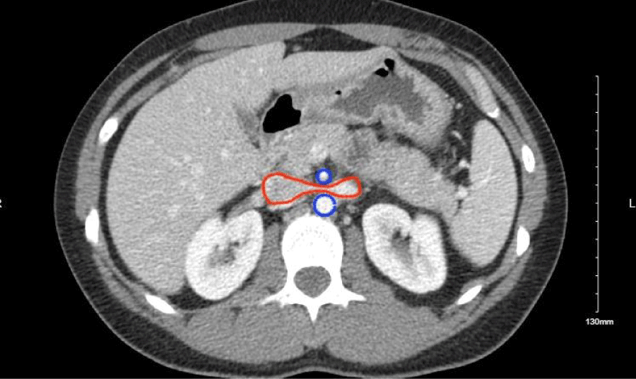 Figure 1: Patient's CT scan showing the proximal superior mesenteric artery (SMA) off the aorta, both highlighted in blue. The distal SMA compressed the left renal vein, highlighted in red. The marked narrowing of the left renal vein is characteristic of nutcracker syndrome.
View Figure 1
Figure 1: Patient's CT scan showing the proximal superior mesenteric artery (SMA) off the aorta, both highlighted in blue. The distal SMA compressed the left renal vein, highlighted in red. The marked narrowing of the left renal vein is characteristic of nutcracker syndrome.
View Figure 1
Preoperatively, the patient received 1 mg of IV midazolam. Induction was done with 100 mcg of IV fentanyl, 60 mg IV of lidocaine, 150 mg IV of propofol, and 40 mg IV of rocuronium. After, endotracheal intubation was successfully achieved with a MAC 3 blade and 7.0 endotracheal tube. Subsequently, an arterial line in the right radial artery was placed with ultrasound guidance for continuous blood pressure monitoring and blood sampling. The patient's initial vitals showed blood pressure (BP) 103/84 mmHg on the ART line, HR 67 bpm, and SpO 2 100%. 62 minutes into the surgery, the IVC was clamped, and the patient started desaturating with BP 90/46 mmHg, SpO 2 89%, HR 83 bpm. After 200 mcg of phenylephrine was administered and FiO2 increased from 30% to 100 % with manual ventilation, the patient continued to remain hypotensive and hypoxic with BP 70/35 mmHg and SpO 2 at 73%. At this time, the patient was given an additional 200 mcg of phenylephrine, bolused with 500 mL of normal saline, and placed into Trendelenburg position. When this didn’t resolve the issue, the surgery team was asked to unclamp the IVC. With this action, the patient’s vitals began to improve BP 106/54 mmHg on the ART line and SPO 2 of 98%. After adequate resuscitation, the surgery team was able to reclamp the IVC and complete the surgery. Postoperative recovery was closely monitored in the surgical intensive care unit with heparin anticoagulation and strict bed rest. The procedure successfully re-established venous drainage, and the patient reported immediate symptom relief postoperatively.
Differential diagnoses for acute intraoperative hypotension included (1) hemorrhage or surgical blood loss, (2) anesthetic overdose or excessive anesthetic depth, (3) pulmonary embolism or venous air entrainment, and (4) mechanical impedance to venous return from inferior vena cava (IVC) occlusion. The temporal association between IVC clamping and the rapid improvement in hemodynamics following unclamping strongly implicated preload reduction secondary to venous inflow obstruction as the principal etiology. Resuscitative interventions-including manual ventilation to optimize oxygenation, phenylephrine administration, intravenous fluid bolus with normal saline, and Trendelenburg positioning-were effective in restoring venous return and stabilizing circulation. After preload correction, the IVC was successfully re-clamped without recurrence of hemodynamic collapse, allowing surgery to proceed uneventfully. Phenylephrine was selected for its selective α-adrenergic agonism, which increases systemic vascular resistance with minimal effect on heart rate or myocardial oxygen consumption [5,6]. Ephedrine was avoided because its reliance on endogenous catecholamine release and cardiac output–mediated effect renders it less effective in states of impaired venous return. In addition, desaturation ensued as a result of decreased cardiac output and ventilation perfusion mismatch.
The observed hemodynamic instability shares mechanistic similarities with post-reperfusion syndrome (PRS), a phenomenon described primarily in liver transplantation and major vascular surgery [6]. PRS is characterized by an abrupt decrease in mean arterial pressure and systemic vascular resistance following reperfusion of ischemic tissue, leading to transient circulatory collapse. Although the hypotensive episode in this case preceded reperfusion, both conditions involve abrupt disruption of venous return and intravascular volume redistribution, compounded by myocardial strain and autonomic imbalance.
Patients with Nutcracker Syndrome and concomitant Pelvic Congestion Syndrome may be particularly vulnerable to such hemodynamic disturbances [1,2]. Chronic pelvic venous hypertension leads to the dilation and remodeling of collateral venous pathways, most notably the gonadal and pelvic veins, resulting in reduced venous compliance and capacitance. Over time, this adaptation diminishes the venous system’s ability to buffer intravascular volume fluctuations. Consequently, even transient IVC clamping may precipitate an exaggerated reduction in preload, with downstream effects on stroke volume and systemic perfusion.
Chronic venous hypertension may therefore conceal underlying circulatory fragility until challenged by open vascular intervention. While endovascular stenting is associated with fewer acute hemodynamic disturbances, surgical transposition remains a definitive treatment option in select cases, albeit with increased anesthetic and perioperative complexity [3,4,7]. Contemporary systematic review of 578 patients reported symptom resolution rates of 92% for LRV transposition, though with the highest reintervention rate at 28.5% [7]. Case series data demonstrated that LRV transposition improved symptoms in 10 of 11 patients with NCS [8].
Management of such patients requires proactive anesthetic planning with invasive hemodynamic monitoring, aggressive volume resuscitation, and readiness for vasoactive support [9]. Positional maneuvers, such as Trendelenburg positioning, may play an important role in augmenting venous return during IVC manipulation. In this case, early recognition and prompt intervention facilitated rapid hemodynamic recovery and an uncomplicated postoperative course.
This case highlights the importance of dynamic and individualized anesthetic strategies in patients with venous congestion syndromes undergoing central vascular surgery. It also raises the possibility of an underrecognized vascular phenotype in which chronic venous pathology impairs circulatory adaptability. Further investigation is warranted to elucidate the relationship between pelvic venous disorders and intraoperative hemodynamic instability, to optimize perioperative management and improve outcomes in this unique patient population.
Our institution does not require ethical approval for reporting individual case.
We obtained written informed consent from the patient to present this case.
Not applicable.
The authors declare that they have no competing interests.
Not applicable.
JL and SC wrote the manuscript. DAF and CL helped to design the case report and revised the manuscript. All authors reviewed and approved the final draft.
Not applicable.