
Aortic dissection, a life-threatening condition, is characterized by a tear in the inner layer of the aorta, allowing blood to surge between the layers, forcing them apart. Type B Aortic Dissection, explicitly impacting the descending aorta, can present with various complications that magnify its severity [1]. A notable concern is the formation of a haemothorax, where blood accumulates in the pleural space, severely compromising respiratory function [2]. Additionally, a false lumen, a secondary channel formed due to the dissection, can result in sluggish blood flow and potential leaks. Levy (2023) [3]. If not addressed, these leaks can escalate into endoleaks post-endovascular interventions, further complicating the clinical scenario. While surgical and endovascular interventions offer potential solutions, the intricate nature of aortic dissection requires a meticulous approach to prevent and manage associated complications. Such complications' severity and potential fatality underscore the critical importance of early diagnosis, intervention, and multidisciplinary management.
A 46-year-old male with a known history of hypertension was admitted following complaints of severe back pain and an episode of syncope. Initial investigations using an urgent CT Aortogram identified a Left haemothorax, a complicated Type B Aortic Dissection, and a contained lea.
On the first day of his admission, an emergency intervention involving a Thoracic Endovascular Aortic Rupture (TEVAR) procedure was carried out-the procedure utilized Cook devices to cover the left subclavian artery while preserving the coeliac artery axis. In the intervention, an angiogram was employed to identify the true lumen, followed by the embolization of the left subclavian artery origin using Nestor coils, specifically of sizes 20mm and 14mm.
Despite the intervention above, a post-procedure assessment showed a sluggish flow within the patent false lumen. This suggested a potential leak originating from the proximal region of the descending aorta, even after the stent had been fully expanded.
Consequently, after holding multidisciplinary discussions and considering the patient's situation, the decision was made to perform an urgent Total Arch Replacement, combined with a frozen elephant trunk (FET) technique, on the second day of his admission.
Postoperatively, the patient was meticulously monitored in the intensive care unit. However, on the 14 th day post-admission, he developed a complete white-out of the left haemothorax. This necessitated another urgent CT Aortogram, which revealed the persistence of the false lumen without any evident contrast leak.
In response, the patient underwent an urgent Left video-assisted thoracoscopy surgery. This surgical intervention revealed the presence of bloody pleural fluid, though active bleeding was not detected.
Further consultations led to the decision to undertake another TEVAR procedure on the patient's 21 st day of admission. This intricate procedure encompassed the embolization of the false lumen combined with in-situ Laser Fenestration. Critical steps in the procedure involved cannulating and pre-stenting the Celiac artery, placing a catheter within the false lumen for subsequent coil insertions, and deploying a tapered TX2 dissection stent. This stenting approach was notably aggressive, intentionally shuttering the SMA by 50%. This was subsequently remedied by positioning an 11mm x 29 VBX stent protruding into the aorta. Large coils were introduced into the false lumen, followed by a stenting procedure on the celiac artery after laser fenestration. The procedure culminated with a completion angiography, which confirmed satisfactory outcomes.
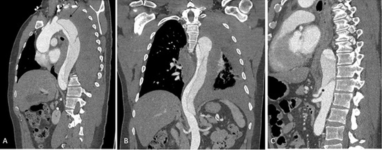 Figure 1: CT aortography in complex type B aortic dissection.
Oblique sagittal CT reformat (A) shows complex dissection flap distal to the origin of left subclavian artery, with the smaller true lumen pushed to the anteromedial aspect of the arch of aorta and descending thoracic aorta by the larger false lumen, which shows faint contrast opacification.
Note the left hemothorax (attenuation 45 HU) and the irregular contour of the false lumen superiorly (black arrow), suggesting ruptured dissection.
Coronal oblique (B) and sagittal oblique (C) CT reformats show the distal extent of the dissection, reaching just above the celiac axis origin (asterisk).
View Figure 1
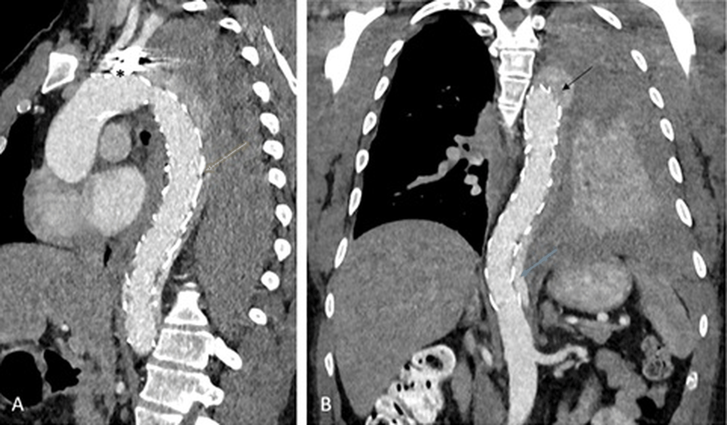 Figure 2: CT aortography after TEVAR.
Sagittal oblique CT reformat (A) shows TEVAR stent in situ (yellow arrow) and the coiled left subclavian artery (asterisk). There is faint contrast opacification of the false lumen, which is better appreciated on the coronal oblique reformat (B), at the level of distal descending thoracic aorta (blue arrow) and distal arch (black arrow) with thrombosed false lumen in between, suspicious for endoleaks at proximal and distal end of the stent. Residual hemothorax with atelectasis of the left lung is also noted.
View Figure 2
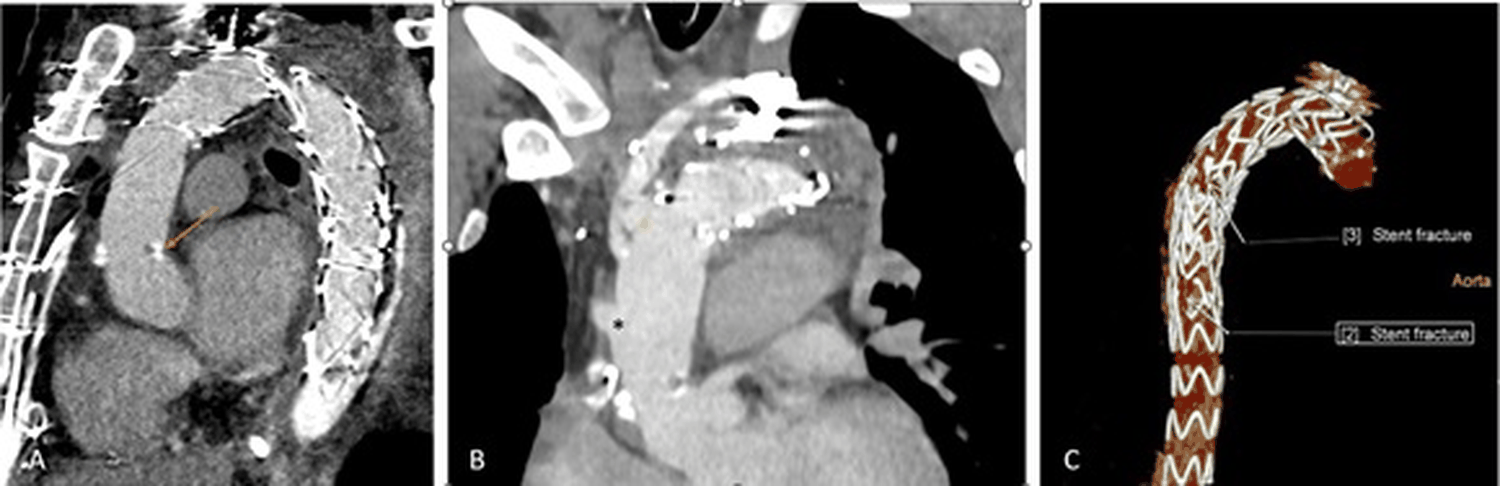 Figure 3: CT aortography after ascending aorta and total arch replacement with frozen elephant trunk surgery.
Sagittal oblique CT reformat (A) depicts the proximal anastomosis site of ascending aorta (orange arrow), without any para-anastomotic leak. Note the persistent endoleak posterior to the TEVAR distally. The endoleak seen previously at the level of the arch is not visualised. The re-implanted brachiocephalic, left common carotid, and left subclavian arterial grafts (indicated by black, white and yellow asterisks respectively) are well opacified as seen on the coronal oblique reformat image (B). 3D reconstruction image (C) shows stent fractures at 2 sites (arrows) in the TEVAR stent.
View Figure 3
Figure 3: CT aortography after ascending aorta and total arch replacement with frozen elephant trunk surgery.
Sagittal oblique CT reformat (A) depicts the proximal anastomosis site of ascending aorta (orange arrow), without any para-anastomotic leak. Note the persistent endoleak posterior to the TEVAR distally. The endoleak seen previously at the level of the arch is not visualised. The re-implanted brachiocephalic, left common carotid, and left subclavian arterial grafts (indicated by black, white and yellow asterisks respectively) are well opacified as seen on the coronal oblique reformat image (B). 3D reconstruction image (C) shows stent fractures at 2 sites (arrows) in the TEVAR stent.
View Figure 3
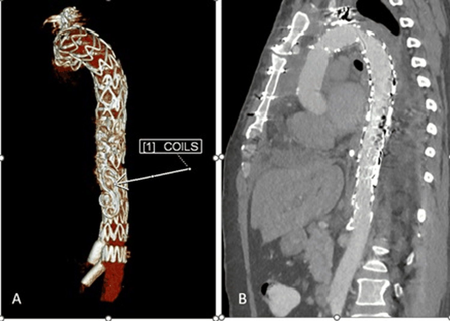 Figure 4: CT aortography post endoleak repair.
3D reconstructed image (A) shows the coils along the posterolateral aspect of the TEVAR stent used to occlude the endoleaks. There is extension of the stent inferiorly with stenting of the celiac axis and SMA as well. Sagittal oblique reformat (B) does not show any opacification of the false lumen, indicating successful management of the endoleaks.
View Figure 4
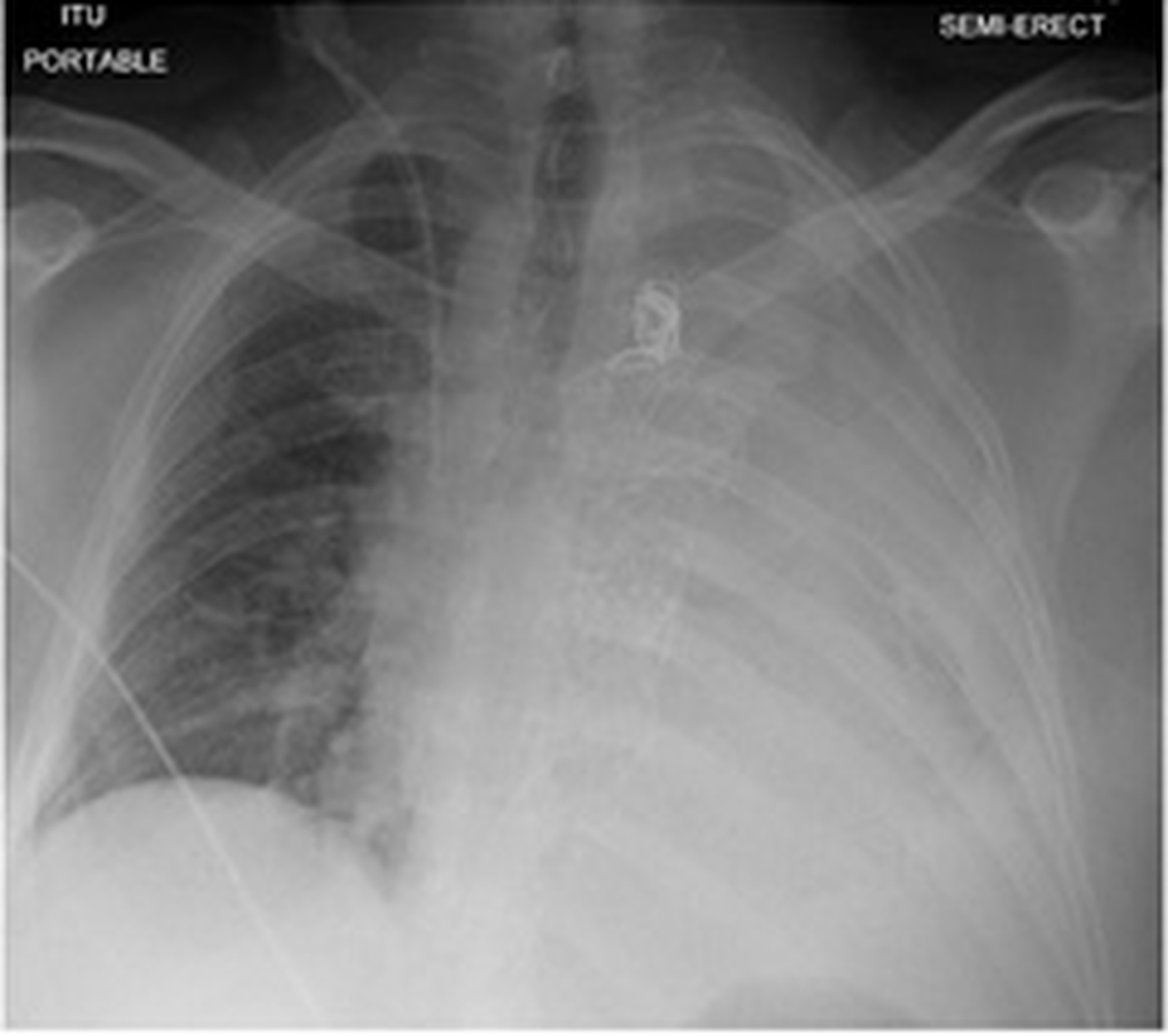 Figure 5: Chest radiograph done after the TEVAR depicts the stent and the coils in left subclavian artery in situ. The left opaque hemothorax is in keeping with left pleural effusion/hemothorax.
View Figure 5
Figure 5: Chest radiograph done after the TEVAR depicts the stent and the coils in left subclavian artery in situ. The left opaque hemothorax is in keeping with left pleural effusion/hemothorax.
View Figure 5
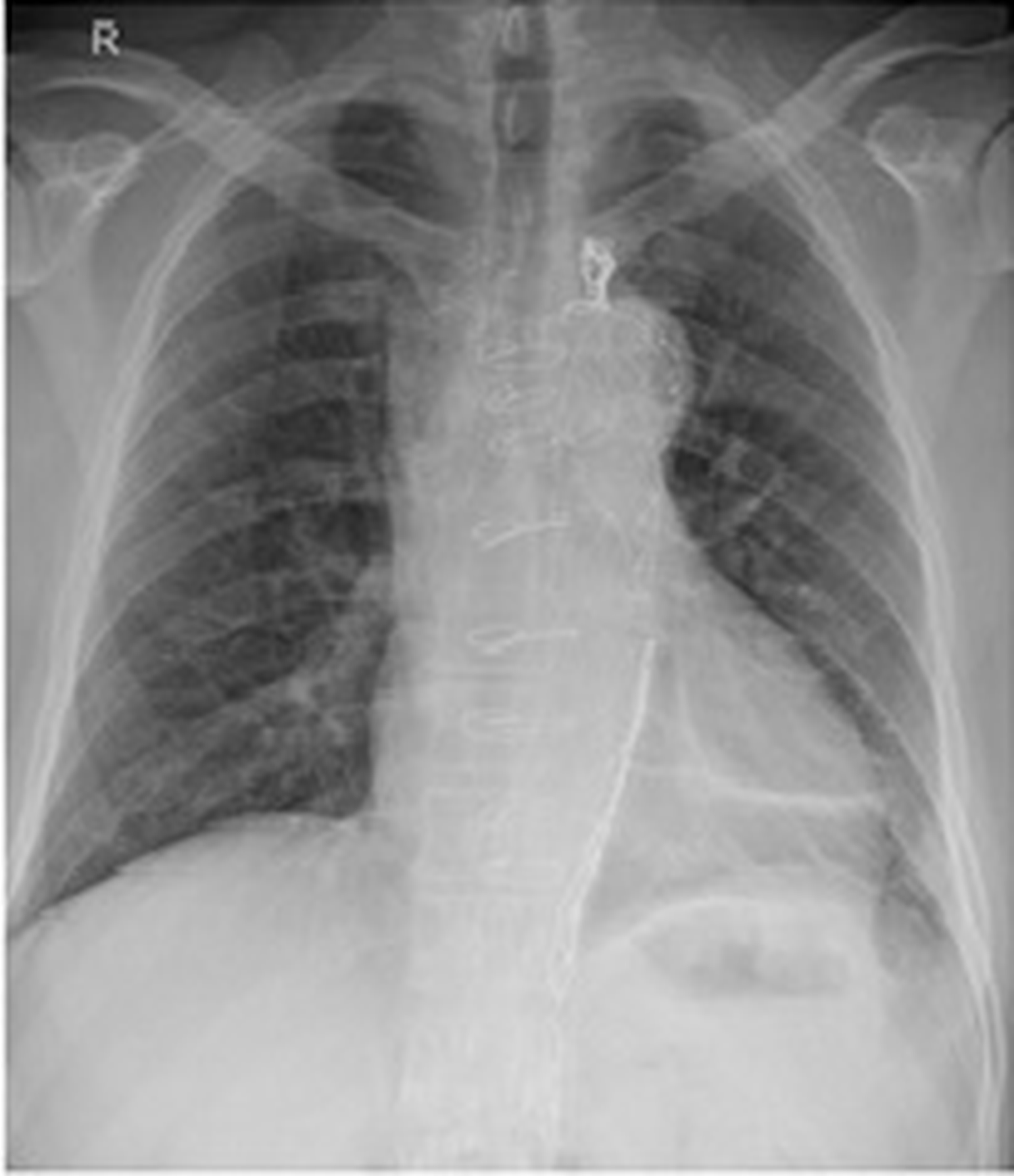 Figure 6: Follow-up chest radiograph taken nearly 2 months after the completion surgery reveals resolution of the left hemothorax with minimal subsegmental atelectasis in the left lung base. Sternotomy wires and coils along the lateral aspect of TEVAR stent are noted.
View Figure 6
Figure 6: Follow-up chest radiograph taken nearly 2 months after the completion surgery reveals resolution of the left hemothorax with minimal subsegmental atelectasis in the left lung base. Sternotomy wires and coils along the lateral aspect of TEVAR stent are noted.
View Figure 6
Follow-up investigations, including a repeat CT Aortogram, showcased the successful resolution of the endoleak. The patient underwent continued rehabilitation within the hospital premises and was eventually discharged on the 45th-day post-admission.
Subsequent outpatient department reviews and a chest X-ray (CXR) confirmed the resolution of the haemothorax.
Acute type B aortic dissection, comprising roughly one-third of all aortic dissection cases, is a condition with a historical legacy dating back centuries. [4] However, progress in understanding its optimal diagnosis and treatment has been slower, particularly in the latter half of the 20 th century. In response, the International Registry of Acute Aortic Dissection (IRAD) was established in 1996 [5]. IRAD has played a pivotal role in advancing our understanding of aortic dissection. Recent analyses of IRAD data have explored temporal classification, risk stratification, and the role of thoracic endovascular aortic repair (TEVAR) in improving outcomes. In 2014, research indicated that the incidence rate, currently at 14 per 100,000 persons per year, up to 30% are type B dissections. [4] research indicated that it has a morbidity rate of 3 in 100,000 people. According to McClure, et al., 2018, [6] 60% of people do not show any signs of rupture malperfusion [7].
Aortic dissection can arise from various factors. Predominantly, hypertension is observed in 70% of patients with Stanford-type TBAD. An abrupt rise in blood pressure from strenuous activities or drugs like cocaine and ecstasy is also notable. Genetic conditions, such as Marfan syndrome, Ehlers-Danlos syndrome, and Turner syndrome, play significant roles. [8] Marfan syndrome notably leads to cystic medial necrosis [9]. Other factors include pre-existing aortic aneurysm, atherosclerosis, complications during pregnancy (especially with conditions like Marfan syndrome), [10] familial predispositions, prior aortic interventions, and certain infectious or inflammatory diseases [3].
Aortic dissection's incidence is 5-30 cases per 1 million annually. While age is a risk factor, most dissections occur between 50 and 65. Differences are observed in older vs. younger patients with ascending aorta dissections: older ones often have atherosclerosis or prior aortic issues, whereas younger patients are linked to conditions like Marfan syndrome. Typically, AAD is thrice as common in men, but women show worse outcomes when affected [3].
Hypertensive individuals, like our 46-year-old patient, are at a heightened risk, underlining the crucial role of blood pressure management in forestalling such severe complications. The heightened vulnerability of hypertensive patients emphasizes the broader public health importance of blood pressure control and regular monitoring. The patient's presentation with severe back pain and syncope was indicative and typical of aortic dissection, and the decision to proceed with an urgent CT Aortogram proved pivotal. Delayed diagnosis in such cases can lead to increased morbidity and even mortality. In this case, prompt detection not only allowed for swift intervention but likely played a role in the eventual positive outcome.
The 2023 systematic review and meta-analysis by Zhao et al. investigated the optimal timing for endovascular repair in acute versus subacute uncomplicated Type B Aortic Dissection. Analysing data from six studies comprising 3,769 patients, it was found that the acute group (2,642 patients) exhibited significantly higher risks of 30-day complications (OR = 1.51), 30-day mortality (OR = 2.39), and 1-year mortality (OR = 1.71) compared to the subacute group (1,127 patients). Reintervention was also more likely in the acute group (OR = 1.42). No significant differences were observed in long-term mortality. These results suggest that subacute endovascular repair may be preferable due to lower risks, but further research with larger cohorts and longer follow-up is needed for confirmation [7].
In conclusion, managing a complicated Type B Aortic Dissection in our 46-year-old hypertensive male patient exemplifies the intricate nature of this cardiovascular condition. This case underscores several crucial points in the clinical management of aortic dissections. First and foremost, early diagnosis is paramount. Identifying the condition through an urgent CT Aortogram enabled rapid intervention, likely pivotal in the patient's eventual positive outcome. Our case highlights the importance of tailored and multidisciplinary approaches. The complexity of aortic dissection necessitated a cascade of interventions, including Thoracic Endovascular Aortic Rupture (TEVAR), Total Arch Replacement with a frozen elephant trunk (FET) technique, and further TEVAR with in-situ Laser Fenestration. Each decision was made in collaboration with various medical specialists, demonstrating the significance of teamwork and expertise in managing such challenging cases. Persistent complications and the subsequent successful resolution of the endoleak and haemothorax emphasize the need for meticulous post-procedure monitoring and follow-up. This case offers valuable insights into managing complicated Type B Aortic Dissection. It serves as a reminder of the critical importance of timely diagnosis, multidisciplinary decision-making, and tailored interventions in optimizing patient outcomes. It contributes to the ongoing research and clinical understanding of aortic dissections, ultimately improving patient care in cardiovascular medicine.
The team acknowledges the contributions of all healthcare professionals involved in the patient's care, emphasizing the multidisciplinary approach critical for successfully managing this case.