
Boerhaave's syndrome is a rare spontaneous oesophageal perforation. It is classically caused by forceful emesis and retching.
We report a 16-month-old boy who presented to the emergency department because of externalized blood from his mouth after his meal. Initial examination was unremarkable. His hemoglobin was at 10 g/dL. Hours later, he developed sudden massive upper gastrointestinal bleeding leading to hemorrhagic shock and cardiac arrest. Post-mortem examination revealed an aorto-oesophageal fistula complicating an oesophageal rupture with mediastinitis. This case highlights an exceptional and fatal presentation of oesophageal perforation in early childhood and emphasizes the need for early recognition of Boerhaave's syndrome.
Boerhaave’s syndrome, Oesophageal perforation, Aorto-oesophageal fistula, Pediatric Gastrointestinal bleeding
BS: Boerhaave's syndrome; CT Scan: Computed Tomography Scan
Boerhaave’s syndrome (BS) is a surgical emergency defined as a spontaneous perforation of the oesophagus without external instrumentation and with no other known esophageal diseases.
This syndrome was first described in 1724 by the Dutch physician Herman Boerhaave. It was thought to be caused by the sudden increase in the intraluminal oesophageal pressure associated with the negative intrathoracic pressure after forceful vomiting or retching [1].
Oesophageal perforation is a fatal condition associated with high mortality rates (more than 50%). BS is the second most common cause of oesophageal perforation after iatrogenic causes. It accounts for 15% of all causes [2].
Clinical symptoms depend on the site of the perforation. Patients may present with chest, neck or abdominal pain, odynophagia, dysphagia, dysphonia, respiratory distress and hematemesis. Complications include mediastinitis, pleural effusion, peritonitis, and septic or hemorrhagic shock.
Due to its atypical presentation, diagnosis of BS is frequently delayed and is often misdiagnosed initially. Early recognition and timely management are strongly associated with improved outcomes. Owing to its rarity, there are no standardized treatment guidelines [3], and management ranges from conservative therapy to aggressive surgical interventions, including oesophagectomy [1].
We present the case of a 16-month-old boy who was admitted to the emergency department for blood externalization occurring after a meal. Ten days earlier, he had been diagnosed with gastroenteritis, during which he experienced several episodes of vomiting and weight loss. He was treated symptomatically. He was a healthy child, with no underlying comorbidities; his vaccinations were up to date, and there was no significant family history. There was no history of foreign body ingestion.
Upon arrival, clinically, he was active but pale. The remainder of the clinical examination was normal. Laboratory investigations showed mild anemia with normal coagulation indices. His chest X-ray was unremarkable. In view of his symptoms, he was admitted for observation, and no specific treatment was initiated at that stage.
Approximately four hours after his evening meal, the patient’s condition deteriorated abruptly. He became agitated, inconsolable, and subsequently vomited blood. He was transferred to the intensive surveillance unit, a second peripheral venous line was inserted, and a blood cell count control was done. Within minutes, he progressed to a cardiac arrest. The patient was intubated, and cardiopulmonary resuscitation was initiated. A continuous blood transfusion and volume expansion with 0.9% saline solution were administered. Vasopressors were administered according to the protocol. Approximately 500 mL of blood was aspirated from his stomach. Unfortunately, we were unable to achieve a return of spontaneous circulation.
No diagnostic investigations or surgical interventions were possible due to the rapid deterioration and progression of his symptoms.
Autopsy revealed two longitudinal oesophageal ruptures, each approximately 1 cm in length, located in the lower third of the oesophagus, associated with mediastinitis. One of these ruptures communicated with the aortic arch, forming an aorto-oesophageal fistula (Figure 1, 2 and 3).
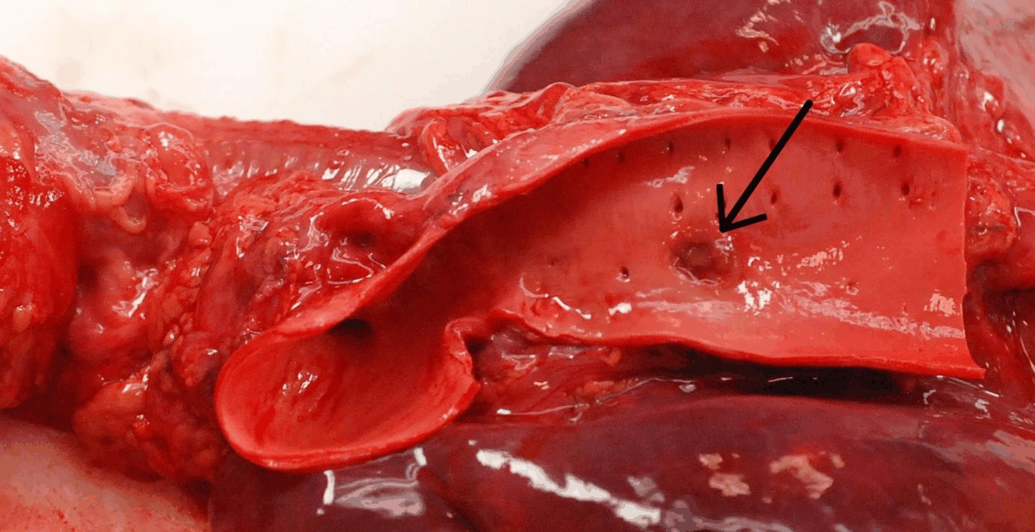 Figure 1: Site of aortic perforation.
View Figure 1
Figure 1: Site of aortic perforation.
View Figure 1
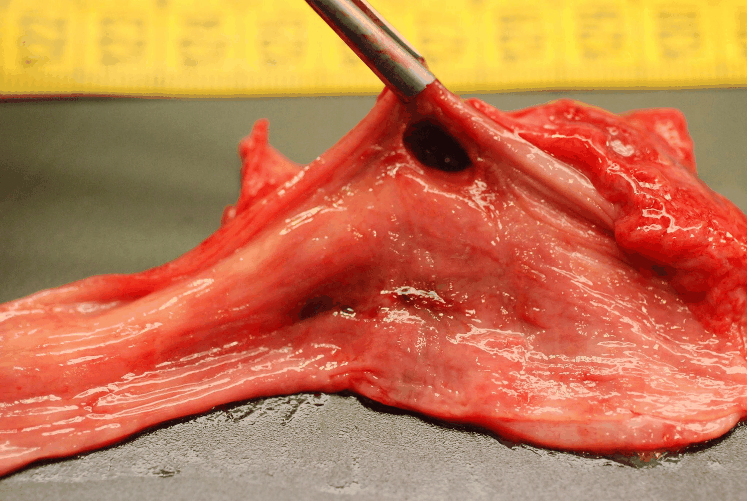 Figure 2: Site of the oesophageal perforation.
View Figure 2
Figure 2: Site of the oesophageal perforation.
View Figure 2
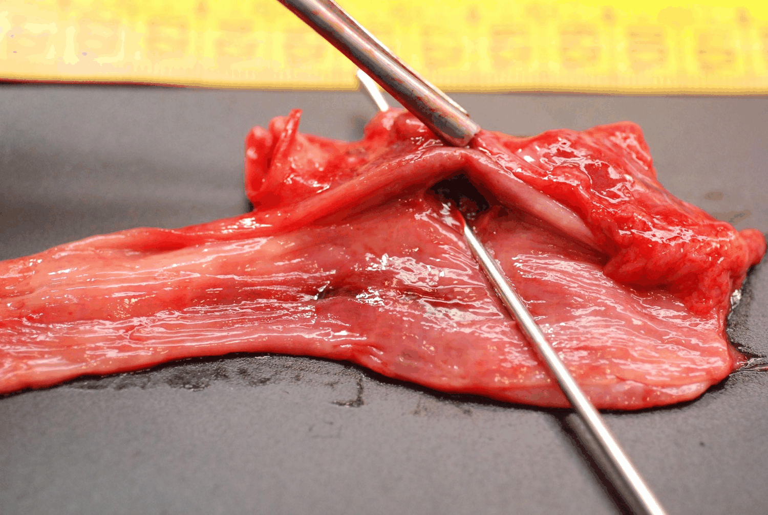 Figure 3: The aorto-oesophagus fistula.
View Figure 3
Figure 3: The aorto-oesophagus fistula.
View Figure 3
We conclude that the massive upper gastrointestinal bleeding resulted from an aorto-oesophageal fistula secondary to BS, leading to severe hemorrhagic shock and cardiac arrest.
Boerhaave’s syndrome is defined by a spontaneous rupture of the oesophagus following forceful vomiting or retching. It accounts for 15% of oesophageal ruptures and is frequently reported in the adult population and rarely in children [1].
Symptoms are often nonspecific, and they can be more challenging to identify in children, especially toddlers, who are unable to report discomfort. These symptoms are frequently associated with a history of emesis or retching.
The typical clinical presentation, Mackler triad (vomiting, chest pain, and subcutaneous emphysema), is uncommon, which can delay the diagnosis and early intervention [4]. Wilson, et al. reported that in patients diagnosed with BS, 25-45% did not present with vomiting before the beginning of the symptoms [5].
About 90% of the spontaneous perforations are located in the distal part of the esophagus, with the majority in the left posterolateral aspect of the intrathoracic oesophagus, possibly due to an anatomical weakness at that point [6]. However, in very young patients, the oesophagus usually perforates into the right pleural cavity [4].
The esophageal rupture may present with various complications, such as mediastinitis or peritonitis, and may lead to sepsis with multiple organ failure [7].
In our case, the patient presented with haematemesis and no other systemic symptoms. The oesophagus rupture got complicated with mediastinitis. The inflammation caused an aortitis, then a tunnelling of an aorto-oesophageal fistula. The patient did not present any fever or discomfort before the hemorrhage.
The prognosis depends mainly on the time between the injury and diagnosis, the location and severity of the rupture, the patient's age, medical history, and finally, the patient's status upon first presentation [8]. Delayed diagnosis and appropriate treatment within the first 12 to 24 hours enhance the good prognosis with a survival rate approaching 75% [1,4].
If not treated in time, a high mortality rate is reported within these patients, ranging between 20% to 40% [8].
In some cases of esophageal rupture, treatment may be conservative (antibiotics and percutaneous drainage of collections) or surgical [4]. The different treatment consensus reported in the literature depended on the rupture location, the team's experience and treatment options.
In our case, the diagnosis was based on autopsy findings. Due to the severity of the hemorrhage, our patient deteriorated rapidly and went into cardiac arrest. The question we asked afterwards was about interest in using a Sengstaken-Blackmore tube. Unfortunately, we don’t have a specific answer.
We report this case to highlight the possibility of an oesophageal rupture in children with an unknown history and after a gastroenteritis episode. Symptoms are discreet and nonspecific to the complication. The indication of a CT scan is in question in our case, with the absence of further clinical symptoms of mediastinitis. A late result of blood culture showed the presence of Streptococcus pyogenes, a week after the death. This result may reflect the history of mediastinitis.
The management of patients with BS is a case-by-case approach; diagnosis and treatment management depend on the patient’s clinical presentation and symptom history.
Boerhaave’s syndrome is a real diagnosis and therapeutic challenge. Assembling different symptoms to orient the diagnosis is a must. We concluded from our case that even children can be victims of this spontaneous rupture. The diagnosis may be more challenging in the pediatric population than in adults. Prognosis depends on an early diagnosis and treatment.
The authors declare that no human subjects were involved in this research. We employed the patient data collection protocols of our work centre for the databases. No personal data were published in this article that could identify the patient, and so informed consent was not required. This study meets the current bioethical research regulations.
No financial support was received in relation to this article.
The authors declare that there is no conflict of interest.