
• Persistent refractory hypokalaemia in oncology patients should not be attributed to gastrointestinal losses alone, even in the presence of stomas or malabsorption.
• Severity, renal potassium wasting, and resistance to replacement are critical triggers for early endocrine investigation.
• Ectopic ACTH secretion may present without classical Cushingoid features and can manifest predominantly as acute metabolic instability.
• Early recognition and initiation of medical adrenal blockade can be life-saving and may stabilize patients sufficiently for onward oncological or palliative care.
Ectopic adrenocorticotropic hormone (ACTH) secretion is an uncommon but potentially life-threatening paraneoplastic syndrome; most often associated with pulmonary neuroendocrine tumours. Gastrointestinal malignancies represent a rare source and may be diagnostically challenging, particularly in the absence of classical phenotypic features of Cushing’s syndrome.
We report the case of a 68-year-old woman with metastatic small bowel adenocarcinoma who presented with severe, persistent hypokalaemia, hypocalcaemia and hypomagnesaemia identified incidentally during routine pre-chemotherapy assessment. Despite aggressive intravenous replacement and repeated hospital admissions, electrolyte abnormalities remained refractory. The disproportionate severity of metabolic derangement and evidence of renal potassium wasting prompted endocrine evaluation, which revealed marked ACTH-dependent hypercortisolism with failure of dexamethasone suppression. Pituitary imaging was normal. Histopathological re-review of tumour tissue demonstrated neuroendocrine differentiation, supporting a diagnosis of ectopic ACTH secretion.
Initiation of medical adrenal blockade with metyrapone, alongside physiological glucocorticoid replacement, resulted in rapid biochemical stabilization and clinical improvement. This case highlights the importance of recognising ectopic ACTH secretion as a cause of refractory dyselectrolytaemia in oncology patients and underscores the need for early endocrine involvement when standard explanations fail.
Ectopic ACTH secretion accounts for approximately 10-20% of cases of ACTH-dependent Cushing’s syndrome and is most commonly associated with pulmonary neuroendocrine tumours and small-cell lung carcinoma. Gastrointestinal sources are rare and frequently under-recognized, particularly when clinical features of hypercortisolism are absent or overshadowed by comorbid disease.
In acute and general medical settings, electrolyte disturbances in patients with cancer are commonly attributed to gastrointestinal loss, chemotherapy-related effects, or poor oral intake. Failure to recognize hormonally driven causes can result in repeated admissions, delayed diagnosis and significant morbidity. This case illustrates how ectopic ACTH secretion may present primarily as severe metabolic instability, posing a diagnostic challenge for non-endocrinologists.
A 68-year-old woman was referred from oncology outpatient services following routine pre-chemotherapy blood tests that revealed profound hypokalaemia (2.3 mmol/L), hypocalcaemia (1.7 mmol/L) and hypomagnesaemia (0.59 mmol/L). Her medical history included Crohn’s disease, chronic kidney disease stage 3, hypertension and a total proctocolectomy with permanent ileostomy performed in 1993. She had recently been diagnosed with small bowel adenocarcinoma with extensive hepatic metastases and a suspected osseous lesion.
She reported progressive fatigue and intermittent muscle cramps but denied increased stoma output, diarrhoea, vomiting or recent weight change. Examination demonstrated proximal muscle weakness but no overt Cushingoid features such as facial rounding, striae or truncal obesity.
Initial management focused on presumed gastrointestinal electrolyte loss, with aggressive intravenous replacement during multiple admissions. Despite escalating supplementation, abnormalities persisted. During one admission, she developed symptomatic hypokalaemia requiring high-dose replacement via central venous access and was transferred to the intensive care unit for cardiac monitoring.
The severity, persistence and resistance to replacement of electrolyte abnormalities prompted reconsideration of the initial diagnostic assumption.
Table 1: Endocrine investigations demonstrated marked ACTH-dependent hypercortisolism
Table 1: Endocrine investigations demonstrated marked ACTH-dependent hypercortisolism. View Table 1
Low-dose dexamethasone suppression testing failed to suppress cortisol levels, supporting ACTH-dependent hypercortisolism. Magnetic resonance imaging of the pituitary demonstrated normal morphology with no evidence of adenoma (Figure 1).
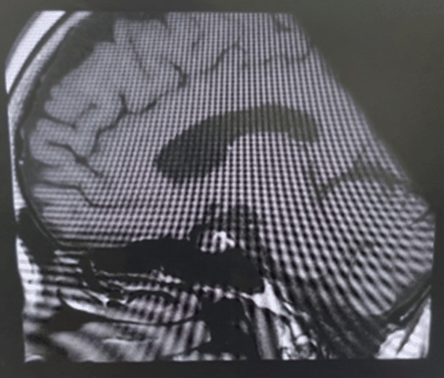 Figure 1: Magnetic resonance imaging of the pituitary gland demonstrating normal morphology with no evidence of adenoma.
View Figure 1
Figure 1: Magnetic resonance imaging of the pituitary gland demonstrating normal morphology with no evidence of adenoma.
View Figure 1
Whole-body positron emission tomography demonstrated multiple fluorodeoxyglucose-avid hepatic lesions consistent with metastatic disease (Figure 2). Histopathological re-review of tumour tissue demonstrated adenocarcinoma with immunohistochemical features of neuroendocrine differentiation. Although direct ACTH immunostaining was not available, the convergent biochemical, radiological and clinical findings supported a diagnosis of ectopic ACTH secretion.
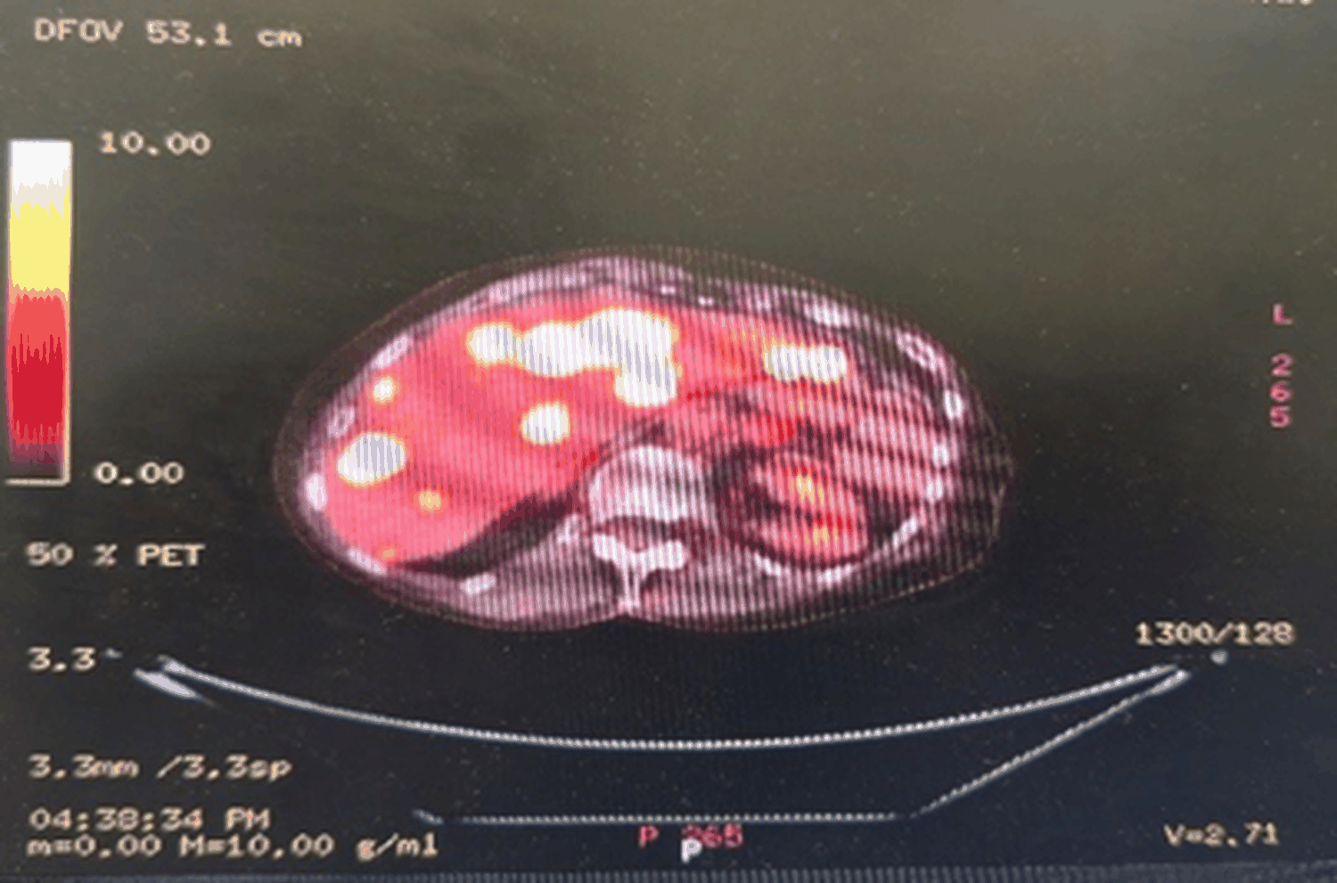 Figure 2: Positron emission tomography scan demonstrating multiple fluorodeoxyglucose-avid hepatic lesions consistent with metastatic disease.
View Figure 2
Figure 2: Positron emission tomography scan demonstrating multiple fluorodeoxyglucose-avid hepatic lesions consistent with metastatic disease.
View Figure 2
The patient was managed in the intensive care unit with continuous cardiac monitoring and aggressive electrolyte replacement. Medical adrenal blockade was initiated using metyrapone (750 mg three times daily), with concurrent physiological hydrocortisone replacement to prevent adrenal insufficiency.
Following treatment initiation, serum cortisol levels declined and electrolyte abnormalities stabilized progressively. Muscle cramps resolved and the patient’s functional status improved. She was discussed at a regional neuroendocrine tumour multidisciplinary meeting, where the diagnosis of ectopic ACTH secretion secondary to neuroendocrine differentiation within metastatic small bowel adenocarcinoma was agreed.
Given advanced disease, management was palliative. Capecitabine was commenced but discontinued due to treatment-related confusion. Long-acting octreotide was introduced to address persistent neuroendocrine activity. The patient was discharged with sustained biochemical stability under joint endocrine and oncology follow-up.
Electrolyte disturbance is a frequent trigger for acute medical admission in patients with malignancy and is commonly attributed to gastrointestinal loss, renal impairment, or treatment-related effects. While these explanations are often valid, diagnostic anchoring to a pre-existing condition can delay recognition of alternative and potentially life-threatening causes when abnormalities are severe, recurrent, or resistant to replacement.
In this case, the presence of a permanent ileostomy and a history of inflammatory bowel disease provided a plausible explanation for hypokalaemia. However, several features were inconsistent with gastrointestinal loss alone: the severity of hypokalaemia, its persistence despite aggressive intravenous replacement, objective evidence of renal potassium wasting, and the need for critical care admission for cardiac monitoring. These features should prompt early diagnostic reassessment rather than repeated empirical replacement.
Ectopic ACTH secretion represents an uncommon but important cause of acute metabolic instability. Unlike pituitary-dependent Cushing’s disease, it frequently presents without classical phenotypic features and may manifest predominantly through biochemical derangement. Excess cortisol activates renal mineralocorticoid receptors, leading to renal potassium wasting, metabolic alkalosis, and sodium retention. Concomitant hypomagnesaemia further impairs potassium repletion, rendering replacement strategies ineffective. For the general physician, recognition of this biochemical pattern is often more informative than reliance on physical features of hypercortisolism, which may be absent or masked by comorbidity.
Diagnosis of ectopic ACTH secretion in patients with advanced malignancy is challenging and often delayed. While tumour immunohistochemistry for ACTH is the diagnostic gold standard, it is not always available or feasible in routine practice. In such circumstances, diagnosis relies on convergent evidence: marked ACTH-dependent hypercortisolism, failure of dexamethasone suppression, absence of pituitary pathology on imaging, compatible tumour histology, and rapid biochemical response to steroidogenesis inhibition. In this case, the consistency of these findings provided sufficient diagnostic confidence to guide management.
Early initiation of medical adrenal blockade is critical. Metyrapone can rapidly reduce cortisol excess, stabilize electrolytes, and improve clinical status, even when definitive oncological treatment is not possible. Prompt endocrine involvement once hormonally mediated electrolyte disturbance is suspected may prevent repeated admissions, escalation of care, and potentially fatal complications.
From a general medical perspective, this case highlights the importance of pattern recognition over attribution. Severe hypokalaemia with renal potassium wasting, resistance to supplementation, or recurrent admissions should act as diagnostic triggers for endocrine investigation, regardless of the presence or absence of classical features of Cushing’s syndrome.
In patients with malignancy, persistent or refractory electrolyte disturbance should prompt diagnostic reassessment rather than repeated empirical replacement. Severe hypokalaemia that is resistant to supplementation or associated with renal potassium wasting should raise suspicion of hormonally mediated causes.
Ectopic ACTH secretion may present predominantly as acute metabolic instability and can be easily overlooked outside specialist settings. Early recognition and initiation of medical adrenal blockade can prevent life-threatening complications and may stabilize patients sufficiently to allow appropriate oncological or palliative care. For general and acute physicians, recognising when electrolyte abnormalities do not fit common patterns is critical to timely diagnosis and improved outcomes [1-7].
Written informed consent was obtained for publication.
The author(s) declared no potential conflicts of interest with respect to the research, authorship, and/or publication of this article.
The authors received no financial support for the research, authorship, and/or publication of this article. The research was performed as part of the employment of the authors in QEQM Hospital
Mohamed Hamza wrote the article, Tamer Mohamed Zaalouk and Abu Bocus shared in the discussion and with Siraj Nasim in collecting the data and revision of the manuscript.