
A 70-year-old male with a history of bilateral surgically treated inguinal hernias, presents with a one-day history of abdominal pain, nausea and vomiting, abdominal distension and obstipation. On examination, the patient was found in good general condition, had a pulse rate of 90 bpm, a blood pressure of 120/60 mmHg, and a temperature of 37°C.
Plain abdominal radiograph (Figure 1) shows dilated loops of small bowel with gas-fluid levels. CT scan (Figure 2) shows proximal and distal small bowel obstruction with a transitional zone in the right lower abdomen. Beyond the transitional zone, there was a cluster of bowel loops within a hernia sac occupying the right supra-vesical fossa behind the abdominal wall, suggesting a supra-vesical internal hernia. There was some fluid within the hernia sac, and the bowel wall enhancement was preserved.
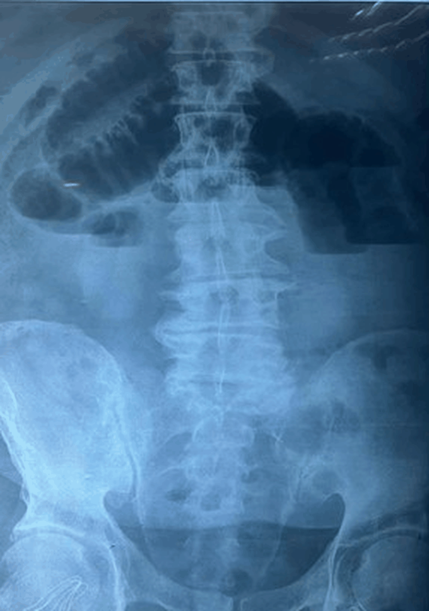 Figure 1: Plain abdominal radiograph showing dilated loops of small bowel with gas-fluid levels.
View Figure 1
Figure 1: Plain abdominal radiograph showing dilated loops of small bowel with gas-fluid levels.
View Figure 1
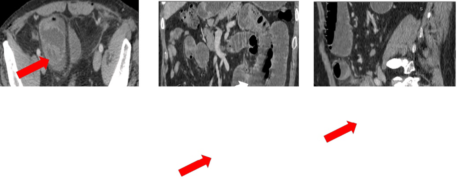 Figure 2: CT scan images on axial (A), coronal (B) and sagittal (C) planes showing proximal and distal small bowel obstruction (White arrows) with a transitional zone in the right lower abdomen with a cluster of bowel loops (Red arrows) within a hernia sac occupying the right supra-vesical fossa.
View Figure 2
Figure 2: CT scan images on axial (A), coronal (B) and sagittal (C) planes showing proximal and distal small bowel obstruction (White arrows) with a transitional zone in the right lower abdomen with a cluster of bowel loops (Red arrows) within a hernia sac occupying the right supra-vesical fossa.
View Figure 2
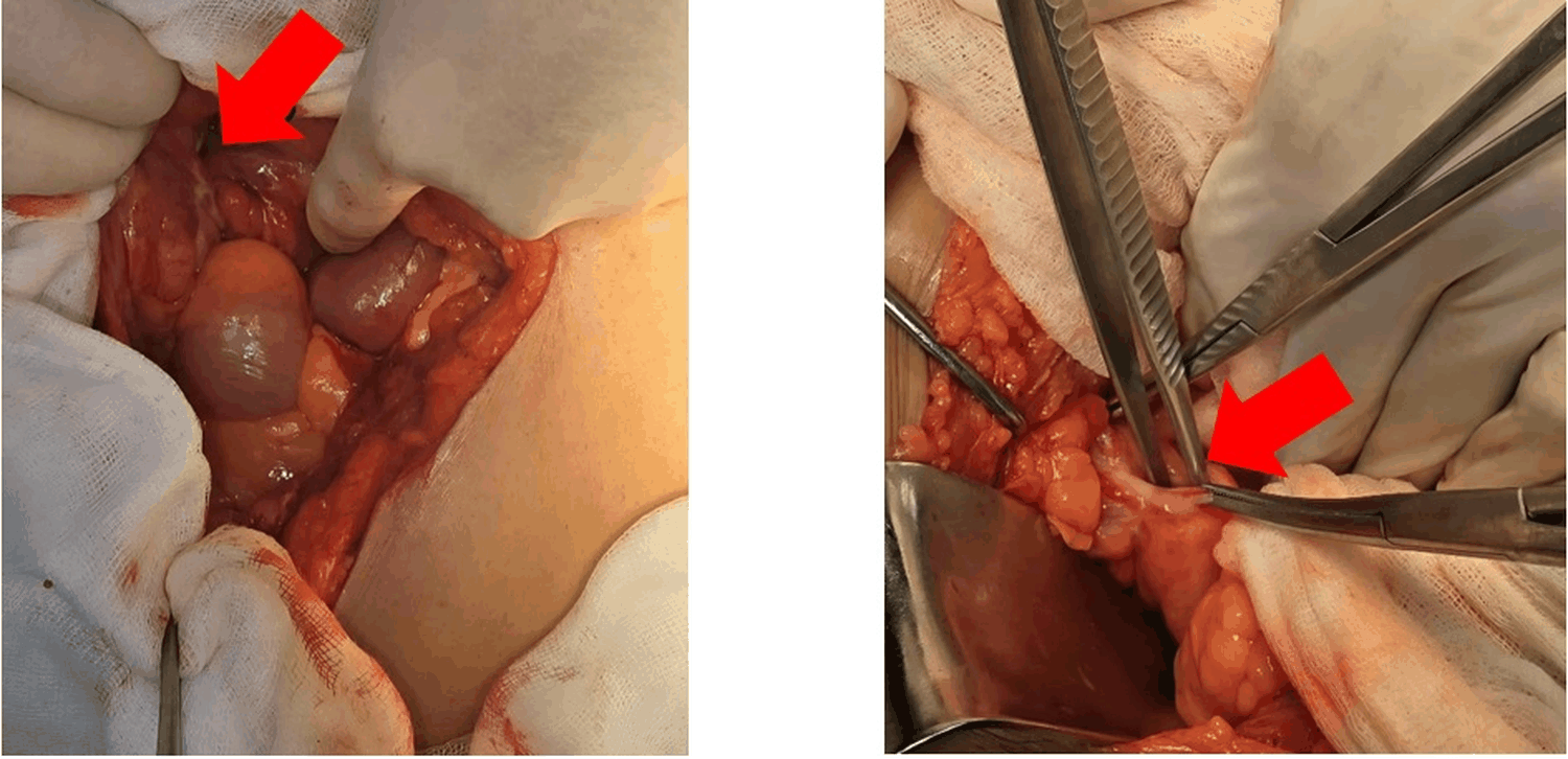 Figure 3: Per-operative images revealing a loop of ileum herniating through a defect (Red arrow) in the right supra-vesical fossa.
View Figure 3
Figure 3: Per-operative images revealing a loop of ileum herniating through a defect (Red arrow) in the right supra-vesical fossa.
View Figure 3
Internal hernias are a rare etiology of small bowel obstruction (SBO), amongst which supra vesical hernia in particular has an extremely low incidence with fewer than a hundred reported cases in the literature [1]. It occurs when a cluster of small bowels is incarcerated in the supra vesical fossa [2], an anatomic space located between the umbilical arteries and the remnants of the urachus. Detecting this variant can be challenging to make on CT scans and it is not rare for the diagnosis to be made preoperatively, increasing the risk of bowel ischemia [1,3,4].
The usual clinical presentation includes nonspecific symptoms of small-bowel obstruction, including distension, obstipation, abdominal pain and vomiting [1]. Urinary symptoms, albeit rare, are possible due to the bladder being compressed by the hernia sac [3]. With no specific signs, suspicion is usually low leading to the possibility of the diagnosis being delayed [1,3]. Surgical intervention is mandatory for reduction of the herniated bowel, assessment of viability, and closure of the hernial defect [2,5].
Plain radiographs may help identify predominantly central dilated loops with gas-fluid level, a sign suggesting small bowel obstruction, without any indicator to the underlying etiology.
Computed tomography (CT), is the imaging modality of choice for suspected internal hernia [1,2]. For internal supra-vesical hernias, characteristic CT features include:
• Sings of closed-loop obstruction, with at least two transition points located near the bladder level with a cluster of small-bowel loops situated in the supra-vesical fossa [1,3].
• Displacement of the bladder [3].
• CT is also useful for appreciating signs of ischemia: bowel wall thickening, reduced or absent enhancement, pneumatosis or mesenteric congestion [3].
Multiplanar reformats (MPR) are helpful in appreciating the disposition of the herniated loops vis à vis the bladder as well as identifying the hernial defect [1]. Recognition of this entity on imaging is crucial to aid in early surgical planning, avoiding complications [2,4,5].
Exploratory laparotomy was performed, revealing a loop of ileum herniating through a pouch in the right supra-vesical fossa. The hernia defect was about 2cm. (Figure 3)
The incarcerated bowel was viable and the hernial defect was closed after reduction.
Post-operative period was uneventful.
The patient was discharged on day 7.
Take Home Message/Teaching Points
Although being an extremely rare cause of small bowel obstruction, radiologists must be familiar with the appearance of supra-vesical hernias and suspicion should be raised when visualizing a sac-like mass of clustered small bowels occupying the supra-vesical fossa, prompting urgent surgical intervention.
Supra-vesical hernia causing small bowel obstruction.
• Closed loop obstruction due to an adhesion
• Small bowel volvulus